Reinforcing Effect of a Cyanoacrylate Adhesive on Surgical Suture Knots
Reinforcing Effect of a Cyanoacrylate Adhesive on Surgical Suture Knots
Folder:
Year:
Abstract:
Despite the latest polymer materials and surgical suturing techniques, the knot will always be the weakest point of the tied suture loop. In theory, the knot must be as small as possible to prevent an excessive amount of tissue reaction and a delay in healing. There have been reports suggesting that topical cyanoacrylate adhesives could have a reinforcing effect on a surgeon’s knot. Such an outcome could lead to the elimination of knot slippage and the unsatisfactory performance of some surgical knots. The main purpose of this study was to determine if the cyanoacrylate adhesive could have a significant reinforcing effect on typical suture types and sizes when tied as a surgeon’s knot. The second aim was to evaluate if the cyanoacrylate adhesive could replace an additional throw in the surgeon’s knot so as to achieve an equivalent mechanical performance. The topical cyanoacrylate adhesive LiquiBand® was combined with six different suture materials (TicronTM, SurgidacTM, Ethilon*, Nurolon*, BiosynTM and PDS*II) in four different sizes (USP 5-0, USP 3-0, USP 0 and USP 1). The surgeon’s knot (2=1) with and without one (2=1=1) and two additional throws (2=1=1=1) were tied in a reproducible way and mechanically tested. Six dependent variables were used to evaluate the performance of each knot with and without adhesive. The performance criteria were: the force at loop failure, the maximum loop holding force, the loop holding capacity, the knot efficiency, the knot elongation efficiency and the loop distraction. From the results and from scanning electron microscopic observations, the cyanoacrylate adhesive was found to significantly improve the knot performance. The improvement was superior with braided sutures and with absorbable polymer sutures. The reinforcement was more significant with thicker suture sizes and with the plain surgeon’s knot. However, one cannot conclude that the improvement created by the adhesive equaled the improvement obtained by the addition of an extra throw for most suture types.
Type of document:
Language:
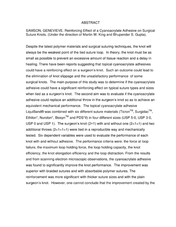
ABSTRACT
SAMSON, GENEVIEVE. Reinforcing Effect of a Cyanoacrylate Adhesive on Surgical
Suture Knots. (Under the direction of Martin W. King and Bhupender S. Gupta).
Despite the latest polymer materials and surgical suturing techniques, the knot will
always be the weakest point of the tied suture loop. In theory, the knot must be as
small as possible to prevent an excessive amount of tissue reaction and a delay in
healing. There have been reports suggesting that topical cyanoacrylate adhesives
could have a reinforcing effect on a surgeon’s knot. Such an outcome could lead to
the elimination of knot slippage and the unsatisfactory performance of some
surgical knots. The main purpose of this study was to determine if the cyanoacrylate
adhesive could have a significant reinforcing effect on typical suture types and sizes
when tied as a surgeon’s knot. The second aim was to evaluate if the cyanoacrylate
adhesive could replace an additional throw in the surgeon’s knot so as to achieve an
equivalent mechanical performance. The topical cyanoacrylate adhesive
LiquiBand® was combined with six different suture materials (TicronTM, SurgidacTM,
Ethilon*, Nurolon*, BiosynTM and PDS*II) in four different sizes (USP 5-0, USP 3-0,
USP 0 and USP 1). The surgeon’s knot (2=1) with and without one (2=1=1) and two
additional throws (2=1=1=1) were tied in a reproducible way and mechanically
tested. Six dependent variables were used to evaluate the performance of each
knot with and without adhesive. The performance criteria were: the force at loop
failure, the maximum loop holding force, the loop holding capacity, the knot
efficiency, the knot elongation efficiency and the loop distraction. From the results
and from scanning electron microscopic observations, the cyanoacrylate adhesive
was found to significantly improve the knot performance. The improvement was
superior with braided sutures and with absorbable polymer sutures. The
reinforcement was more significant with thicker suture sizes and with the plain
surgeon’s knot. However, one cannot conclude that the improvement created by the
adhesive equaled the improvement obtained by the addition of an extra throw for
most suture types.
Reinforcing Effect of a Cyanoacrylate Adhesive
on Surgical Suture Knots
by
Genevieve Samson
A thesis submitted to the Graduate Faculty of
North Carolina State University
in partial fulfillment of the
requirements for the Degree of
Master of Science
Textile Engineering
Raleigh, North Carolina
2009
APPROVED BY:
_______________________________
Tushar K. Ghosh
______________________________
Hechmi Hamouda
________________________________
Martin W. King
Co-Chair of Advisory Committee
________________________________
Bhupender S. Gupta
Co-Chair of Advisory Committee
Dedication
What matters is not how much you know but what you do with it.
ii
Biography
Genevieve Samson was born in Vancouver, Canada, while still a young child her
family moved to Quebec City, Canada where she earned her Bachelor of
Engineering degree in Mechanical Engineering from Laval University in 2006. As a
young graduate, she was eager to learn more and to focus her attention on the
textile industry, thus she found a project manager’s position in a nonwoven
company, located in Quebec, Canada. In pursuit of further studies, she challenged
herself to research fiber and textile science abroad. She was accepted to North
Carolina State University, the leading textile school in the United States, for a
master’s program in Textile Engineering in Fall 2007. The focus of her research has
been the mechanical properties of surgical sutures. She expects to graduate in
May, 2009 and return to the textile industry.
iii
Acknowledgments
I would like to start by expressing my appreciation to my co-advisors, Dr. Martin W.
King and Dr. B.S. Gupta. To Dr. King, a deep and sincere thank you for your
guidance, but more importantly for providing me with two of the most valuable things
in my life: education and travel experiences. Thank you for giving me these life
changing opportunities. To Dr. Gupta, thank you for your wise counsel and the
opportunity to work on the knot security project. I also wish to give my special
thanks to other members of my committee: Dr. Tushar K. Ghosh and Dr. Hechmi
Hamouda. In addition, I would like to thanks Dr. Stephen Michielsen for his
instruction on fracture mechanics theory.
This project would not be possible without the support of the College of Veterinary
Medicine.
I would like to thank in particular Dr. Simon Roe for the use of his
laboratory and equipment; also to Dr. Kyle Mathews for his guidance and assistance
in acquiring suture materials.
I shall not forget John Hash for his time and
availability.
Special thanks to Advanced Medical Solutions Group plc. for their quick and
enthusiastic support and for providing the Liquiband® samples. Thanks to Ethicon,
Inc., more specifically to Mr. Patrick Terry for providing the suture samples used in
this study and Mr. Joe Hotter from Covidien for providing suture samples as well.
Your participation was much appreciated.
iv
From my heart, I would like to express my sincere appreciation to all the members of
the Biomedical Textile Laboratory at the College of Textiles. Joshua, thank you for
your good ear; Sarah, thank you for your strength and Nilesh, thank you for your
help and continuous assistance.
Finally, I would like to thank my love for his sacrifices, for his help and incessant
support through this entire odyssey. Yoakim, thank you ten thousand times over for
always believing in me and my craziest projects.
v
Table of Contents
List of Tables ............................................................................................................. x
List of Figures .......................................................................................................... xiii
1. Introduction ............................................................................................................ 1
1.1 Problem Statement .......................................................................................... 2
1.2 Goals and Objectives ....................................................................................... 2
1.3 Limitations ....................................................................................................... 3
2. Review of Literature .............................................................................................. 5
2.1 Surgical Sutures ............................................................................................... 5
2.2 Knot Definition .................................................................................................. 7
2.3 Types of Surgical Knots ................................................................................... 8
2.4 Knot Challenges and Limitations ................................................................... 10
2.5 Knot Performance .......................................................................................... 11
2.5.1 Knot Mechanics ........................................................................................ 11
2.5.2 Type of Knot Failure ................................................................................. 12
2.5.3 Surgical Knot Evaluation .......................................................................... 13
2.6 Tissue Adhesives ........................................................................................... 15
2.6.1 Cyanoacrylate Adhesive .......................................................................... 15
2.6.1.1 History of Cyanoacrylate Adhesive ................................................... 15
2.6.1.2 Cyanoacrylate Chemistry ................................................................. 16
vi
2.6.1.3 Utilization ........................................................................................... 17
2.6.1.4 Cyanoacrylate Toxicity ..................................................................... 18
2.6.2 Fibrin Glue ............................................................................................... 19
2.6.3 Other Adhesives ....................................................................................... 20
2.7 Prior Art ......................................................................................................... 20
3. Materials and Methods ......................................................................................... 23
3.1 Independent Variables ................................................................................... 23
3.2 Dependent Variables ..................................................................................... 24
3.3 Definitions ...................................................................................................... 24
3.4 Design of Experiment .................................................................................... 28
3.5 Materials ........................................................................................................ 29
3.6 Methods ......................................................................................................... 33
3.6.1. Specimen Preparation ............................................................................ 33
3.6.2 Linear Density and Suture Diameter ....................................................... 33
3.6.3 Breaking Force and Elongation ............................................................... 34
3.6.4 Tying Tension .......................................................................................... 35
3.6.5 Loop Tying .............................................................................................. 37
3.6.5.1 Knot Formation, Step by Step Procedure ......................................... 39
3.6.6 Loop Testing ............................................................................................ 41
3.6.6.1 Loop Testing, Step by Step Procedure ............................................. 41
3.6.7 Specimen Analysis .................................................................................. 42
3.6.8 Data Analysis .......................................................................................... 42
3.6.9 Statistical Analysis of Loop Performance ................................................ 44
vii
3.6.9.1 Normality Test .................................................................................. 44
3.6.9.2 Variance Test ................................................................................... 44
3.6.9.3 Mean Test ........................................................................................ 44
4. Results and Discussion ....................................................................................... 46
4.1 Results ............................................................................................................ 46
4.1.1 Results Obtained on Straight Suture ........................................................ 46
4.1.2 Results Obtained on Suture Loop ............................................................ 47
4.1.2.1 Examples of Force-Elongation Curves ............................................. 47
4.1.2.2 Plots of Mechanical Performance of Suture Loop ............................ 49
4.1.2.3 SEM Pictures ................................................................................... 64
4.2 Discussion ..................................................................................................... 66
4.2.1 General Objective .................................................................................... 66
4.2.2 Specific Objectives .................................................................................. 66
4.2.2.1 Effect of Suture Material .................................................................... 67
4.2.2.2 Effect of Suture Structure .................................................................. 70
4.2.2.3 Effect of Suture Coating ................................................................... 71
4.2.2.4 Effect of Suture Size ......................................................................... 72
4.2.2.5 Effect of the Number of Throws ........................................................ 75
4.2.2.6 Knot Equivalence ............................................................................. 77
4.2.3 Standard Deviation of TicronTM Suture .................................................... 79
4.2.4 Dependent Variables Evaluation ............................................................. 79
5. Conclusion .......................................................................................................... 80
5.1 Conclusions .................................................................................................... 80
viii
5.2 Recommendations and Future Work .............................................................. 81
6. References .......................................................................................................... 83
7. Appendix .............................................................................................................. 88
7.1 Matlab Program No.1 ..................................................................................... 89
7.2 Matlab Program No. 2 .................................................................................... 91
7.3 Plot of Breaking Force and Elongation .......................................................... 98
7.4 Mean and Standard Deviation of the Dependent Variables for Every Knot Type
............................................................................................................................. 99
7.5 Statistical Analysis of the Performance Criteria ........................................... 104
7.6 ANOVA results for the six specific objectives .............................................. 108
ix
List of Tables
Table 2.1 Commercial suture coating [2] .................................................................. 6
Table 2.2 Suture USP sizes and corresponding diameters [3]................................... 7
Table 2.3: Physical properties of alkyl 2-cyanoacrylates and cured properties [3] ... 16
Table 3.1: Design of experiment ............................................................................. 29
Table 3.2: Suture material used in the study ........................................................... 30
Table 3.3: Digital pictures of the monofilament and braided sutures ...................... 31
Table 3.4: Physical measurements of suture materials (M: monofilament and B:
braided) ................................................................................................................... 34
Table 3.5: Force at loop failure for a 6-throws square knot and the tying tension used
(M: monofilament and B: braided) ........................................................................... 37
Table 4.1: Breaking force and elongation of suture material (M: monofilament and B:
braided).................................................................................................................... 46
Table 4.2: Two-way analysis of variance results for size USP 0 knot 2=1 with and
without adhesive. (No: no adhesive, With: with adhesive, P: PDS*II, E: Ethilon*, T:
TicronTM and B: BiosynTM)2 ..................................................................................... 68
Table 4.3: Two-way analysis of variance results for size USP 3-0 knot 2=1=1 of
Ethilon* (Mono) and Nurolon* (Braided) with and without adhesive. (No: no
adhesive, With: with adhesive) ............................................................................... 71
Table 4.4: Two-way analysis of variance results for BiosynTM, knot type 2=1(No: no
adhesive, With: with adhesive) ............................................................................... 74
Table 4.5: Two-way analysis of variance results for BiosynTM size USP 3-0 (No: No
adhesive, With: With adhesive) ............................................................................... 76
x
Table 4.6: Two-way analysis of variance results comparing all suture material of size
USP 0 for 2=1 knot with adhesive (w) and 2=1=1 knot (B: BiosynTM, E: Ethilon*, P:
PDS*II and T: TicronTM) ........................................................................................... 78
Table 7.1: Dependent variable results for all sutures for 2=1 knot .......................... 99
Table 7.2: Dependent variable results for all sutures for the 2=1 knot with adhesive
............................................................................................................................... 100
Table 7.3: Dependent variable results for all sutures for the 2=1=1 knot .............. 101
Table 7.4: Dependent variable results for all sutures for the 2=1=1 knot with
adhesive ............................................................................................................... 102
Table 7.5: Dependent variables results for all sutures for 2=1=1=1 knot with and
without adhesive ................................................................................................... 103
Table 7.6: P-values of the Shapiro-Wilk W test for the dependent variables named
Force at loop failure, Maximum loop-holding force and Loop holding capacity ..... 104
Table 7.7: P-values of the Shapiro-Wilk W test for the dependent variables named
Knot efficiency, Knot elongation efficiency and Loop distraction ........................... 105
Table 7.8: P-values of the Bartlett’s Test, Welsh’s T Test and One Way ANOVA test
if applicable for the dependent variable named Force at loop failure, Maximum loopholding force and Loop holding capacity ................................................................ 106
Table 7.9: P-values of the Bartlett’s Test, Welsh’s T Test and One Way ANOVA test
if applicable for the dependent variable named Knot efficiency, Knot elongation
efficiency and Loop distraction ............................................................................... 107
Table 7.10 P-values generated by ANOVA indicating the influence of adhesive and
material on three suture sizes and five materials ................................................... 108
Table 7.11 P-values generated by ANOVA indicating the influence of coating and
adhesive on knot type 2=1 and 2=1=1 .................................................................. 108
xi
Table 7.12 P-values generated by ANOVA indicating the influence of suture size and
adhesive on knot type 2=1 and 2=1=1 for Ethilon*, PDS*II, Ticron TM and BiosynTM
............................................................................................................................... 109
Table 7.13 P-values generated by ANOVA indicating the influence of the number of
throws and the adhesive on Ethilon*, PDS*II, TicronTM and BiosynTM of size USP 3-0
............................................................................................................................... 110
Table 7.14 P-values generated by ANOVA indicating the influence of the material
and the knot type on Ethilon*, PDS*II, TicronTM and BiosynTM .............................. 110
xii
List of Figures
Figure 2.1 : High strength suture material [10] .......................................................... 6
Figure 2.2: Different parts of a surgical knot [12] ...................................................... 8
Figure 2.3: Three major types of surgical knots ........................................................ 9
Figure 2.4: Square knot converted into a slip knot [12] ........................................... 10
Figure 2.5: Duncan loop used in arthroscopy [14] .................................................. 10
Figure 2.6: Knot configuration used to evaluate µ where To is the tension inside the
loop and T1 the tension in the ears [18] .................................................................. 12
Figure 2.7: Testing apparatus with knotted suture loop around 2 aluminum rods
submerged in saline bath [21] ................................................................................. 14
Figure 2.8: Chemical structure of 2-cyanoacrylates where R is the alkyl group ...... 16
Figure 2.9: Application of cyanoacrylate on a clean wound [26] .............................. 17
Figure 2.10: Barbed suture used for wound closure [35] ......................................... 21
Figure 2.11: Knotless device to close a wound [36] ................................................. 21
Figure 2.12: Malleable collar with straight and double loop (a), Lapra-ty® (b) with
suture of size USP 3-0 [37] ...................................................................................... 21
Figure 3.1: Force at loop failure .............................................................................. 25
Figure 3.2: Elongation at loop failure ...................................................................... 25
Figure 3.3: Maximum loop-holding force ................................................................. 26
Figure 3.4: Elongation at maximum loop-holding force ........................................... 26
Figure 3.5: Loop holding capacity ........................................................................... 26
Figure 3.6: Loop distraction .................................................................................... 28
xiii
Figure 3.7: Liquiband® package and ampoules [23] ................................................ 32
Figure 3.8: Straight suture specimen mounted between pneumatics capstan clamps
................................................................................................................................. 32
Figure 3.9: 1=1=1=1=1=1 knot on aluminum mandrel ............................................ 36
Figure 3.10: Tying equipment ................................................................................. 36
Figure 3.11: Surgeon’s knot with additional throws, a. Two throws (2=1), b. Three
throws (2=1=1), c. Four throws (2=1=1=1), d. Five throws (2=1=1=1=1) (6) ........... 38
Figure 3.12: Didactic pictures explaining two steps for the Surgeon’s knot two-hand
tie technique [12]...................................................................................................... 39
Figure 3.13: Knot tying equipment showing the fixed ear and load cell moving in the
direction of the white arrow ..................................................................................... 40
Figure 3.14: Knotted loop specimen mounted between the pins on a tensile testing
machine .................................................................................................................. 41
Figure 4.1: Force-elongation curve for Ethilon* size USP 0 and 2=1 knot .............. 48
Figure 4.2: Force-elongation curve for BiosynTM size USP 5-0 and 2=1=1 knot. ..... 48
Figure 4.3: Performance plots for the BiosynTM suture, size USP 0 with (w) and
without adhesive ...................................................................................................... 50
Figure 4.4: Performance plots for the BiosynTM suture, size USP 3-0 with (w) and
without adhesive ..................................................................................................... 51
Figure 4.5: Performance plots for the BiosynTM suture, size USP 5-0 with (w) and
without adhesive ..................................................................................................... 52
Figure 4.6: Performance plots for the Ethilon* suture, size USP 0 with (w) and
without adhesive ...................................................................................................... 53
Figure 4.7: Performance plots for the Ethilon* suture, size USP 3-0 with (w) and
without adhesive ..................................................................................................... 54
xiv
Figure 4.8: Performance plots for the Ethilon* suture, size USP 5-0 with (w) and
without adhesive ..................................................................................................... 55
Figure 4.9: Performance plots for the Nurolon* suture, size USP 3-0 with (w) and
without adhesive ..................................................................................................... 56
Figure 4.10: Performance plots for the PDS*II suture, size USP 0 with (w) and
without adhesive ...................................................................................................... 57
Figure 4.11: Performance plots for the PDS*II suture, size USP 3-0 with (w) and
without adhesive ..................................................................................................... 58
Figure 4.12: Performance plots for the PDS*II suture, size USP 5-0 with (w) and
without adhesive ..................................................................................................... 59
Figure 4.13: Performance plots for the SurgidacTM suture, size USP 1 with (w) and
without adhesive ..................................................................................................... 60
Figure 4.14: Performance plots for the TicronTM suture, size USP 0 with (w) and
without adhesive ...................................................................................................... 61
Figure 4.15: Performance plots for the TicronTM suture, size USP 3-0 with (w) and
without adhesive ..................................................................................................... 62
Figure 4.16: Performance plots for the TicronTM suture, size USP 5-0 with (w) and
without adhesive ..................................................................................................... 63
Figure 4.17: SEM picture of Ethilon* size USP 5-0, knot 2=1 with adhesive (A:
Adhesive and S: Suture) ......................................................................................... 64
Figure 4.18: SEM picture of PDS*II size USP 5-0, knot 2=1 with adhesive (A:
Adhesive and S: Suture) ......................................................................................... 64
Figure 4.19: SEM picture of TicronTM size USP 3-0, knot 2=1=1 with adhesive (A:
Adhesive and S: Suture) ......................................................................................... 65
xv
Figure 4.20: SEM picture of TicronTM size USP 5-0, knot 2=1 with adhesive (A:
Adhesive and S: Suture) ......................................................................................... 65
Figure 4.21: SEM picture of BiosynTM size USP 5-0, knot 2=1 with adhesive before
(a) and after (b) mechanical test (A: Adhesive and S: Suture) ................................ 65
Figure 4.22: SEM picture of Ethilon* size USP 5-0, knot 2=1 with adhesive before (a)
and after (b) mechanical test (A: Adhesive and S: Suture) ..................................... 66
Figure 4.23: Contact angle θ formed by a drop of liquid on a solid surface ............ 69
Figure 4.24: The low bending rigidity of size USP 5-0 (a) allows a tight knot with not
a lot of room for moisture but it is the opposite for size USP 0 (b) (green: suture,
blue: moisture ......................................................................................................... 73
Figure 7.1: Breaking force of every material used in the study including standard
error bars ................................................................................................................ 99
Figure 7.2: Breaking elongation of every material used in the study including
standard error bars ................................................................................................. 99
xvi
1. Introduction
Sutures have been used for thousands of years in the medical field, and they
continue to be the technique of choice for wound closure, with about 350 million
being used per year in the USA alone [1]. By definition, surgical sutures are sterile
yarns used to hold tissues together until they heal adequately for self-support or they
are used to permanently join tissues with implanted prosthetic devices [2].
Currently, there is an abundance of different materials and methods available to
close wounds, including staples, adhesives as well as permanent and absorbable
sutures.
Regardless of the latest materials and suturing techniques, physicians
must keep in mind the safety and security of their knots. This is because the tied
knot will always be the weakest point of the suture, with strength reductions varying
from 35% to 95% [3]. Once constructed, the knot must be as small as possible to
prevent an excessive amount of tissue reaction. On the other hand, surgeons will
commonly correct for possible knot slippage and early knot breakage by using a
thicker suture or tying a bulkier complex knot with additional throws. The potential
negative effects of increasing the knot volume include increasing inflammation which
results in delayed wound healing, extending the suturing time and the length of the
operation, and/or the development of a suture reaction or fistula. Accordingly, it is
important that an alternative way of improving knot security is found, while at the
same time limiting or diminishing the knot volume.
However, the concept of what “knot security” means is difficult to describe and the
literature is full of definitions which differ with the author.
In the past, some
approaches have been studied that involve welding or fusing the structure of the
knot which have relied on the thermoplastic nature of synthetic sutures [4, 5]. The
approach, while successful in increasing knot security in laboratory experiments,
1
suffered difficulties of precision when applied in a clinical setting. More recently, a
study has reported that the addition of cyanoacrylate adhesive to a surgeon’s knot
can increase the breaking strength of a looped suture significantly and reduce the
amount of slippage [6]. This method could potentially be used successfully during
orthopedic surgery where high tensile loads are involved, such as in tendon repair.
1.1 Problem Statement
It is well known that the knot will always be the weakest point in the suture loop and
this is why suture efficiency will invariably depend on the performance of the knot
itself. Additional throws or thicker sutures can make the knot safer, more secure and
have a reduced risk of slipping but to the detriment of other properties. A recent
study has demonstrated that combining conventional material of size USP 2 suture
with a cyanoacrylate adhesive significantly reinforced the knot itself without the need
for extra throws [6]. However, this investigation was limited to only one size of
suture, and the heavy size reported is rarely used outside of orthopedic surgery.
Therefore, based on these encouraging results, there is a demand among clinicians
for research to be conducted on a broader range of suture types and sizes [7].
1.2 Goals and Objectives
The goal of this study is to determine if cyanoacrylate adhesive can have a
significant reinforcing effect on the surgeon’s knot when tied with typical suture types
and sizes. The ultimate aim is to identify which factors contribute to knot
reinforcement, and to establish a range of optimal reinforcing conditions.
materials to be studied include permanent sutures, namely polyester (Ticron
TM
The
and
SurgidacTM) and nylon (Ethilon* and Nurolon*), as well as absorbable materials
2
(BiosynTM and PDS*II), combined with the topical cyanoacrylate adhesive
LiquiBand®.
The specific objectives in completing these goals are to determine in detail the effect
of reinforcement on the knot performance1 of a looped suture, and more explicitly:
1) To determine the effect of different suture materials on the knot performance
with and without reinforcement.
2) To determine the effect of the type of suture structure on the knot
performance with and without reinforcement.
3) To determine the effect of suture coating on the knot performance with and
without reinforcement.
4) To determine the effect of the size of the suture on the knot performance with
and without reinforcement.
5) To determine the effect of the number of throws on the performance of the
knot with and without reinforcement.
6) To determine if a small reinforced knot can be as safe and secure as a
regular thicker knot.
1.3 Limitations
The suture samples used in this study were supplied by manufacturers. These were
not randomly selected from a population; rather we relied on the availability of the
type of material chosen. In some cases, the recommended material had already
expired or was soon to expire, according to the notation found on the package.
Moreover, the experimental knots used for this research were tied by the
investigator. This investigator does not have a surgeon’s training and no tying
1
Knot performance is defined in the “Definitions” section of Chapter 3 with an explanation of how this
multi-dimensional variable was measured during this study.
3
experiences other than the one gained during this study. The reliability of the
investigator’s knot tying ability has not been compared with that of an experienced
surgeon.
4
2. Review of Literature
2.1 Surgical Sutures
The surgical suture was one of the first biomaterial devices used by medicine. In
fact, the use of linen by the Egyptians to create a suture thread was reported more
than 4000 years ago [3]. Over the years, new natural and synthetic biomaterials
have been developed to produce sutures with enhanced mechanical properties and
reduced inflammatory reaction. By definition, surgical sutures are sterile filaments
used either to hold tissues together until they have healed adequately for selfsupport, or to join tissues with implanted prosthetic devices [2]. A suture device is
composed of a metal needle, usually made of stainless steel, and a thread. This
second component is more important in terms of biocompatibility because it remains
in the wound to hold the tissues together. The thread can be made of absorbable or
nonabsorbable material.
Nonabsorbable sutures will remain permanently in the
body, and can be made of different synthetic polymers, including polyester, nylon,
polypropylene and ePTFE. On the other hand, absorbable suture materials lose a
significant portion of their mechanical strength over a period of 2 to 3 months [3], or
up to one year for new synthetic monofilaments. The first absorbable sutures were
made of catgut. They have been progressively replaced by synthetic copolymer
materials made by mixing flexible polymer segments with high-strength segments
[8]. Absorbable sutures have received increasing interest over the last few decades
and now represent about 42 % of the total suture market worldwide [8]. In terms of
their physical structure, sutures can be classified as monofilament, multifilament,
twisted and braided. The differences in structure affect the handling properties and
the mechanical behavior of the suture. Recently, high strength performance sutures
have emerged on the market (Figure 2.1) and these sutures combine different
structures such as Supramid® (S Jackson Inc., Alexandria, VA, USA), which is a
5
twisted core of nylon enclosed in a smooth nylon 6 outer shell [9]. It is claimed to
reach higher breaking strength while keeping good handling properties.
Figure 2.1: High strength suture material [10]
Since sutures must pass through various tissues with minimal friction, coatings are
applied to multifilament braids and twisted structures to improve their surface
lubricity. There are a variety of coatings available, some absorbable, some not, and
they depend on the type of suture, the type of polymer and the suture’s trade name
(Table 2.1).
Table 2.1 Commercial suture coatings [2]
Coating
Suture Trade Name
Absorbable
Poloxamer 188 (Pluronic F-68)
Dexon
Calcium stearate and copolymer of
Vicryl
glycolide-lactide
Nonabsorbable
Silicone
silk
Ticron
Surgilon
Wax
Nurolon
Poly(tetramethylene adipate)
Ethibond
fluorocarbon
Tevdek, Ethiflex
6
Type of Polymer
Polyglycolide
Poly(glycolide-L-lactide)
(polyglactin 910)
Silk
Polyester
Polyamide (nylon 66)
Polyamide (nylon 66)
Polyester
Polyester
Finally, sutures can be classified according to their size or diameter. A standard
code was developed, agreed to and published in the United States Pharmacopoeia
(USP) in order to define the sizing system of absorbable and nonabsorbable sutures
(Table 2.2).
However, because this national suture sizing standard is widely
accepted, but not well policed, there is a tendency for manufacturers to produce
commercial suture sizes near the upper size limits or even extend the sizing system
illegally beyond that allowed by the USP [11]. In practice, the surgeon must use the
smallest diameter suture that will hold the wound tissue safely and securely without
breaking.
Table 2.2 Suture USP sizes and corresponding diameters [3]
USP Size
Non synthetic absorbable suture
8-0
7-0
6-0
5-0
4-0
3-0
2-0
0
1
2
3
Nonabsorbable and synthetic
absorbable sutures
Diameter limits (mm)
10-0
9-0
8-0
7-0
6-0
5-0
4-0
3-0
2-0
0
1
2
3
4
0.020-0.029
0.030-0.039
0.040-0.049
0.050-0.069
0.070-0.099
0.100-0.149
0.150-0.199
0.200-0.249
0.250-0.299
0.300-0.399
0.400-0.499
0.500-0.599
0.600-0.699
0.700-0.799
2.2 Knot Definition
A surgical knot is composed of three components (Figure 2.2). First, the loop
created by the knot maintains the approximation of the tissues and provides tension
between the divided wound edges [12]. Secondly, the knot itself is composed of a
certain number of throws that are made one after the other.
7
A single throw is
defined as two threads wrapped around each other so that the angle of wrap equals
360 degrees [13]. A sliding throw is one in which the thread enters and leaves the
throw on the same side [14]. Finally, the ears are the cut ends of the suture. They
provide the insurance that the last throw will not unravel if the loop expands or if the
knot slips. The doctor’s side of the knot is defined as the side of the knot with “ears,”
or the side where tension is applied during tying [12]. The patient’s side is defined
as the side of the knot with the loop.
Figure 2.2: Different parts of a surgical knot [12]
In 1976, a standardized nomenclature was created to describe a knot’s
configuration. The number of wraps in each throw is indicated by an Arabic number,
and the relationship between each throw, being either crossed or parallel, is signified
by the symbols X or =, respectively [12]. The presence of a slip knot is indicated by
the letter S instead of an Arabic numeral and the configuration is detailed by using
symbols // and #.
2.3 Types of Surgical Knots
Various types of knots are used to tie sutures, but the principal ones are the square
knot, the granny knot and the surgeon’s knot. The square knot (1=1) has been
investigated and is reported to be the easiest and most reliable for tying the majority
of suture materials [15]. The reason is that its geometry allows high tension points
8
to be located within the strand where it passes around another strand [13]. It is a
single throw followed by a single throw. The right ear and loop both come out on
either the anterior or posterior side of the knot. The left ear and loop come out
opposite to the right ear and loop (Figure 2.3) [13]. The granny knot (1x1) is not
recommended because of its tendency to slip. However, it may be inadvertently tied
by a considerable proportion of surgeons by incorrectly crossing the strands of a
square knot (Figure 2.3) [3]. The surgeon's knot or friction knot is recommended for
tying a lot of materials such as braided synthetic absorbable sutures, coated Vicryl®,
polyester, nylon and polypropylene sutures.
It is composed of a double throw
followed by a single throw; with the right ear and loop coming out on the same side
of the knot (Figure 2.3). Further single throws can be added on top of the surgeon’s
knot to improve security.
Figure 2.3: Three major types of surgical knots
With the new less invasive surgical techniques, such as laparoscopic surgery, new
types of knots have had to be created (e.g. half-hitch or sliding knots). They are
called sliding knots because they can be “slid” for a certain distance, and then be
locked at the desired position. The square knot (1=1) can easily become a slip knot
(S=S) if the surgeon does not reverse the position of his hands after each throw, or,
if a greater tension is apply to one ear (Figure 2.4) [12]. Finally, some sliding knots
have been developed for specific surgical operations like the Duncan loop or the
9
Overhand loop. They are used extensively in the field of arthroscopy (Figure 2.5)
[14].
Figure 2.4: Square knot converted into a
slip knot [12]
Figure 2.5: Duncan loop used in
arthroscopy [14]
2.4 Knot Challenges and Limitations
The significant advances in materials science and engineering over the past
decades have provided surgeons with a wide and complex range of choices and
approaches to wound closure [3]. A series of research studies has been motivated
by the fact that tying a knot in a suture is associated with considerable challenges
and limitations. First, knot tying requires time and extensive training, especially for
less-invasive surgeries. For some surgical operations, tying knots can take as much
as 50% of the surgeon time [16]. Irrespective of the knot configuration and the
suture material, the inherent weakest link in the surgical suture is the knot, and the
second weakest point is the portion immediately adjacent to the knot. The strength
reduction due to knotting can be as large as 35% to 95% [3].
This decline is
attributable to knot slippage, the mechanical crushing of the suture by surgical
instruments and stress concentrations in the knot itself. The applied tension on the
suture strand is transformed into tensile, bending, compression and shear stresses
on the filaments in the knot. These forces and the shearing action break the filament
at a load lower than the simple tensile breaking load [13]. When a knotted suture
10
fails, the consequences may be disastrous, including wound dehiscence, massive
bleeding, and/or incisional hernia [12]. In order to avoid such complications, the
general surgical practice is to either introduce additional throws or to use a thicker
suture. Once formed inside the body, the larger knot can produce a delay in wound
healing, constrict the blood flow and cause a distortion in the tissue which can lead
to necrosis and/or scar formation [17]. Sutures provoke a significant inflammatory
response, particularly if the knot is larger. For example, an increase in size from
USP 4-0 to USP 2-0 increases the volume of subsequent reaction by 137% to 255%
[3].
2.5 Knot Performance
In 1937, Taylor was the first person in modern times to become interested in the
security of surgical knots [13].
Even now, seven decades later, clinicians and
scientists still debate the most accurate way to evaluate the knot performance of a
suture.
2.5.1 Knot Mechanics
It is the friction between suture filaments that allows the knot to stay tied. In order to
take advantage of this phenomenon, the number of crossing points inside the knot
and the contact angle can be increased. The surface of the suture or the filaments
can also be altered (e.g. braided filament or coating). Each time two suture threads
are in contact, the frictional forces created will oppose the tension applied to the
loop. If the knot is in equilibrium, the frictional forces will equal the tension applied at
the loop ends [13]. It has been shown that the coefficient of friction of a suture (µ)
can be established using the following equation, where n is the number of turns and
β is the angle between the two suture strands (Figure 2.6) [18]:
11
T1 = To e µπnβ
Figure 2.6: Knot configuration used to evaluate µ where To is the tension inside the
loop and T1 the tension in the ears [18]
2.5.2 Types of Knot Failure
According to Thacker, there are three types of knot failure modes. 1) The suture
material can yield, fracture or break (called knot breakage), 2) the knot can slip
(called knot slippage), and 3) the knot can untie from the doctor’s side (called knot
untying) [13].
The first mode is the preferred mode of all three. It will happen when the knot has
been tied under the correct tension and locked properly. The ideal surgical knot is
one that requires the least number of throws to achieve knot breakage behavior.
The tissue in which the suture is implanted can also influence the knot strength or
knot security. In the case of absorbable sutures, a progressive decline in knot
breaking strength is expected after tissue implantation [12].
Knot slippage is required up to a certain level. Ideally, the suture should elongate
under low loads to accommodate any developing wound edema, but return to its
original length after healing and resolution of the edema [12]. However, too much
slippage will cause a separation of the wound edges. If the internal geometry has
not reached equilibrium when tied by the surgeon, further tension will make the knot
12
tighten or “snug down”. Secondly, if the tension applied to the loop is higher than the
frictional forces in the knot, then slippage will occur. Multifilament sutures tend to
slip less than monofilament sutures [3]. The degree of knot slippage can also be
influenced by the coefficient of friction, the suture diameter, the type of knot and the
level of moisture in the wound [12].
Finally, there is a small chance that the knot will untie on the doctor’s side. When
using a stiff material, such as a monofilament, and a thicker suture, the last
unrestrained throw has a tendency to open up and untie. This is the reason why it is
important to leave about 3 mm of suture material to form the ears [12].
2.5.3 Surgical Knot Evaluation
As mentioned earlier, evaluating and rating the performance of surgical knots has
been done in the past using different criteria. However, there are certain concepts
that have been broadly accepted. Chu et al has defined the loop holding capacity
(LHC) as either the force required to break a tied suture loop or alternatively to
provoke slippage of at least 2 mm within the knotted loop [3]. The concepts of knot
failure and knot holding capacity (KHC) are only variants of the same LHC.
A
second definition often used in the literature is the knot efficiency. Depending on the
source, the exact meaning of this concept changes slightly. Chu et al has defined it
as half of the loop holding capacity expressed as a percentage of the breaking
strength of the unknotted suture thread [3]. Others have defined it as the tensile
strength of a knotted suture divided by the tensile strength of the unknotted suture
expressed as a percentage [12]. A third and last important concept is the handling
characteristics of the suture.
Surgeons evaluate the handling characteristics of
sutures by constructing knots using manual and instrumental tying techniques. They
will then select that suture which permits a two-throw knot to be easily advanced or
13
“snugged down” [12]. Another way of evaluating the handling properties is to record
the time [14] or the number of steps [19, 20] required to complete the tying of a
particular knot.
Knot security is not a concept that has a clear and straight forward definition. Some
studies have attempted to define knot security as a collection of characteristics and
they have created new test methods and equipment to evaluate the knots’
performance. Ilahi et al have used a cyclic loading protocol in saline solution to test
knotted loops and record the applied load required to generate a 3 mm separation
as well as the ultimate failure load (Figure 2.7) [21]. Hong et al evaluated the knot
performance of new suture materials by testing the knot pull strength, the knot rundown and the knot security. Here, the knot pull strength was the breaking force
applied between the ears of a knotted suture, and the knot security was the breaking
force applied to a knotted suture inside the loop [22].
Figure 2.7: Testing apparatus with knotted suture loop around 2 aluminum rods
submerged in a saline bath [21]
14
Finally, it is important to note that the only way to identify the type of failure is by
visual observation during testing. No other tests or concepts have been found in the
literature to accurately differentiate knot slippage from knot breakage.
2.6 Tissue Adhesives
Unlike sutures that close wound mechanically, tissue adhesives use chemical
bonds. The ideal adhesive should be safe, biodegradable, effective and easy to use
even in moist tissues.
Nowadays, there are different categories of adhesives
available. According to some estimates it is predicted that as much as 40% of the
global suture/staple market could eventually be accounted for by tissue adhesives
and sealants [23].
2.6.1 Cyanoacrylate Adhesive
2.6.1.1 History of Cyanoacrylate Adhesives
Work with cyanoacrylate adhesives started with Ardis in 1949 followed by clinical
uses in 1965 by Watson and Maguda for tympanic membrane repair [24]. At about
the same time, Krazy Glue was being used in the medical field, although it was
found to have severe histotoxicity. A survey conducted in 1984 on the medical
applications of Krazy Glue in USA demonstrated that 34% of the institutions
contacted had a working knowledge of this adhesive [25]. While the FDA prohibited
its usage, more research was being done to develop fast setting and strong n-butyl
cyanoacrylates that were less toxic (e.g. HistoAcryl (B Braun), Indermil (Vygon), or
LiquiBand (Medlogic)). Then, Closure Medical Inc. developed DermaBond, that was
a slower setting octyl-cyanoacrylate adhesive with more flexibility. It was approved
for external clinical use by the FDA in 1998. More recently, we have seen overseas
development
of
blended
butyl
and
octyl
15
cyanoacrylates
(e.g.
LiquiBand
Laparoscopic (Medlogic)) providing both a fast setting adhesive with a good degree
of flexibility [26].
Recent research has demonstrated that the new family of
cyanoacrylate hemostatic agents (OMNEX, Ethicon), which had previously proven
their safety and efficacy in topical use, are now safe and effective absorbable
surgical sealants for internal use.
2.6.1.2 Cyanoacrylate Chemistry
Cyanoacrylate tissue adhesives are liquid monomers that polymerize in the
presence of moisture on contact with tissue surfaces in an exothermic reaction
creating a strong yet flexible film that bonds the apposed wound edges [27]. The
following chemical structure represents the family of 2-cyanoacrylate monomers
(Figure 2.8) and the various properties of the cured adhesive are listed in Table 2.3.
CN
CH2 = C
O = C- O - R
Figure 2.8: Chemical structure of 2-cyanoacrylates where R is alkyl group
Table 2.3: Physical properties of alkyl 2-cyanoacrylates and cured properties [3]
Cranoacrylate
Structure of Alkyl (R)
Cured bonding to Stainless Steel
Viscosity
(cp)
Set time (min)
Strength (kg/cm )
2
Methyl 2-cyaboacrylate
Ethyl 2-cyanoacrylate (Krazy
Glue)
n-Propyl 2-cyanoacrylate
n-Butyl 2-cyanoacrylate
Isobutyl 2-cyanoacrylate
CH3
CH3CH2
2.2
2.9
Coments go here:
- Log in to post comments