A new method that uses cyanoacrylate tissue adhesive to fill scoring incisions in septal cartilage correction
A new method that uses cyanoacrylate tissue adhesive to fill scoring incisions in septal cartilage correction
Folder:
Journal:
Year:
Abstract:
OBJECTIVES/HYPOTHESIS Numerous methods are used in the correction of deviated septal cartilage. One of these methods is to perform partial-thickness incisions (scoring) on the concave side of the deviated cartilage. In this retrospective report, we present a series of patients who were treated by filling the scoring incision gaps with cyanoacrylate-based tissue adhesives to increase the effectiveness of scoring incisions and to maintain stability of the corrected concave cartilage segments.
STUDY DESIGN A retrospective clinical study presenting a patient group who was treated using a new surgical method for septal deviation.
METHODS Twenty-three patients with septum deviation and nasal deformity underwent surgery with the open rhinoplasty approach. Intra- or extracorporeal scoring incisions were performed on the concave side of the deviated septal cartilage, and cyanoacrylate tissue adhesives were applied to the incisions of the corrected cartilage. After polymerization and hardening of the cyanoacrylate tissue adhesive, the operation continued in the normal manner. Preoperative and postoperative clinical results and computed tomography images of the patients were assessed.
RESULTS With a mean 24-month follow-up, all patients with respiratory complaints related to deviated septum reported improvement in nose breathing. Clinical and radiologic observations showed that the corrected septum was stable in its new position. There were no complications arising from the use of cyanoacrylate.
CONCLUSIONS Cyanoacrylate is an effective, instant, safe method of treatment in correcting deviated septal cartilage with scoring incisions and filling the gaps of the incisions.
DOI:
10.1002/lary.21738
Type of document:
Language:
The Laryngoscope
C
V 2011 The American Laryngological,
Rhinological and Otological Society, Inc.
A New Method That Uses Cyanoacrylate Tissue Adhesive to Fill
Scoring Incisions in Septal Cartilage Correction
_
_
¨
Irfan Ozyazgan, MD; Onurkan Idacı, MD
Objectives/Hypothesis: Numerous methods are used in the correction of deviated septal cartilage. One of these methods
is to perform partial-thickness incisions (scoring) on the concave side of the deviated cartilage. In this retrospective report, we
present a series of patients who were treated by filling the scoring incision gaps with cyanoacrylate-based tissue adhesives to
increase the effectiveness of scoring incisions and to maintain stability of the corrected concave cartilage segments.
Study Design: A retrospective clinical study presenting a patient group who was treated using a new surgical method
for septal deviation.
Methods: Twenty-three patients with septum deviation and nasal deformity underwent surgery with the open rhinoplasty approach. Intra- or extracorporeal scoring incisions were performed on the concave side of the deviated septal cartilage, and cyanoacrylate tissue adhesives were applied to the incisions of the corrected cartilage. After polymerization and
hardening of the cyanoacrylate tissue adhesive, the operation continued in the normal manner. Preoperative and postoperative clinical results and computed tomography images of the patients were assessed.
Results: With a mean 24-month follow-up, all patients with respiratory complaints related to deviated septum reported
improvement in nose breathing. Clinical and radiologic observations showed that the corrected septum was stable in its new
position. There were no complications arising from the use of cyanoacrylate.
Conclusions: Cyanoacrylate is an effective, instant, safe method of treatment in correcting deviated septal cartilage with
scoring incisions and filling the gaps of the incisions.
Key Words: Cyanoacrylate, scoring incision, septal deviation.
Laryngoscope, 121:1164–1172, 2011
INTRODUCTION
Many different treatment modalities are used for
correcting septal cartilage fractures and deviations.
Regardless of which treatment is preferred, one of the
main concerns is to maintain the nasal support of the
septum by preserving the septal cartilage as much as
possible.1 The remaining dorsal and caudal septal cartilage must be at least 8- to 10-mm wide after correction.2
Scoring is a well-known method used for giving shape
to the septal cartilage.3,4 Scoring incisions are partial-thickness incisions made on the concave surface of the cartilage.
Correction of bent septal cartilage by using scoring incisions
and holding it in place for support is a method used in the
treatment of septal deviation.2,5 The efficiency of incisions
depends on the deviation rate of the cartilage and the depth
of the incisions. Deeper incisions cause obtuse bending
angles of the cartilage.3 However, excessively deep incisions
From the Department of Plastic, Reconstructive and Esthetic
Surgery, Erciyes University, Faculty of Medicine, Kayseri, Turkey.
Editor’s Note: This Manuscript was accepted for publication January 11, 2011.
This study was partially presented at the 31st National Plastic,
Reconstructive and Esthetic Surgery Congress, Adana, Turkey, October
17–21, 2009.
The authors have no funding, financial relationships, or conflicts
of interest to disclose.
_
¨
Send correspondence to Dr. Irfan Ozyazgan, Erciyes University Faculty of Medicine, Department of Plastic, Reconstructive and Esthetic Surgery, 38039 Melikgazi, Kayseri, Turkey. E-mail: ozyazgan@erciyes.edu.tr
DOI: 10.1002/lary.21738
Laryngoscope 121: June 2011
1164
could result in damage to the integrity of the cartilage and
weaken it. In contrast, scoring incisions that are not sufficiently deep are inadequate for the preferred correction. In
this patient series report, use of cyanoacrylate (CA)-based
tissue adhesive application to increase the efficiency of scoring incisions and to prevent concavity recurrences in septal
deviation treatment is presented.
MATERIALS AND METHODS
Medical records of patients who were referred to Erciyes
University Faculty of Medicine, Department of Plastic, Reconstructive, and Esthetic Surgery, with septum deviation and
nasal deformity between January 2004 and February 2009 were
retrospectively evaluated; among these, 23 patients (17 men, 6
women) who were treated with scoring incisions and CA tissue
adhesives were analyzed. The mean age of the patients was
43.6 (range, 19–63) years.
In addition to routine preoperative assessments, axial and
coronal computed tomography images were obtained for all
patients. Surgery was performed using the open rhinoplasty
technique with general anesthesia. The surgical technique was
briefly as follows: After a submucosal injection of a local anesthetic solution containing half diluted 10% lidocaine and 1/
100,000 adrenaline, subperiosteal and subchondral dissections
were performed to expose the cartilaginous and bony septum.
The deviated bony septum and crest were resected. Rotated cartilaginous septum in the area of the nasal crest or excessively
thickened, folded septum was also excised. In some patients, if
stabilization of the remaining septal cartilage could not be
maintained in its anatomic position or the connection with bony
structures was deficient or if the cartilaginous septum was
¨
Ozyazgan and _
Idacı: Cyanoacrylate and Scoring Incisions
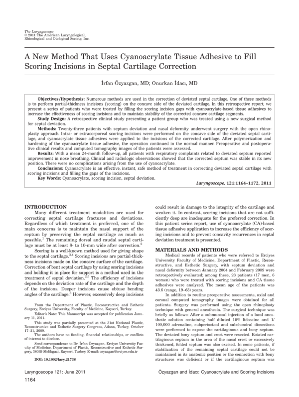
Fig. 1. Upper left, A hingelike deviated septal cartilage to be corrected
extracorporeally; upper right, deviated septal cartilage after scoring in
different directions and the excision
of parts that were not usable or
necessary to the protect septal support; lower left, holding the cartilage
in overcorrected position while waiting for polymerization of the glue;
lower right, the corrected cartilage.
[Color figure can be viewed in the
online issue, which is available at
wileyonlinelibrary.com.]
excessively deformed, the septal cartilage was removed, and the
correction was achieved extracorporeally. In other patients, the
septal cartilage correction procedure was performed in vivo. In
conditions in which the cartilaginous septum was partially
resected, the surgeon paid attention to keep the remaining cartilage as an L strut with at least 10 mm of dorsal septum and
10 mm of caudal septum to maintain the stability of the cartilaginous framework. In aforementioned cases where
manipulations after releasing the septal cartilage from deviated
bony septum were not sufficient to correct the septum or in
those cases in which excessively concave septum (like a thumb
impression) was present, scoring incisions were made and modified by combining CA glue for treatment.
Scoring and CA Usage
Incisions were made with a scalpel to the concave surface
of the septal cartilage, in approximately half the thickness of
the cartilage. The cartilage surface was cleansed of blood and
other liquids before the procedure. The CA adhesive, N-butyl-2cyanoacrylate þ methacryloxysulfolane (Glubran; GEM, Viareggio, Italy) or N-butyl-2-cyanoacrylate (Liquiband; MedLogic Ltd,
Devon, England), was applied to the concave side of the cartilage while holding it in a slightly overcorrected position. The
choice of glues was not made intentionally; they were used
according to availability of hospital resources. GlubranV was
used in the first six patients, and LiquibandV was used in the
remaining 17. When the adhesive was applied to one end of the
incision, the low viscosity of the adhesive made it easy for it to
flow to the other end. After approximately 35 to 40 seconds of
curing time, cartilages were kept untouched to control the
shape and the effectiveness of the procedure. Scoring incisions
R
R
Laryngoscope 121: June 2011
were usually performed in two perpendicular directions because
the majority of these deviations did not occur in a one-way
direction and did not display flexion hinge activity. These deviations appeared like a concavity made by finger pressure and
needed at least two directional multiple scoring incisions. CA
adhesives were carefully applied to the incisions with maximum
attention to prevent them from overflowing. This procedure was
performed to both sides in cases in which undulations had
resulted in concavities in both sides (Figs. 1–6). In addition, in
the last patient in the study, adhesives were scraped off with a
scalpel when overflow was noticed. This procedure did not affect
the shape of the corrected cartilage.
In the extracorporeal correction procedure, cartilages were
put into place and fixed by suturing to both the upper lateral cartilages and to the nasal spine. After the incisions were closed,
silicone intranasal splints with airway passages were placed and
secured with transfixion sutures, and the operation was finished
after nasal packing. Nasal packs were removed 24 hours postoperatively. Intranasal splints were taken out after 2 weeks.
RESULTS
Follow-up interval in these patients ranged from 11
to 62 months (mean follow-up interval was 24 months),
and postoperative computed axial and coronal tomography images of some patients were obtained. One patient
required a rhinoplasty procedure for cosmetic reasons 8
months after the operation. A biopsy was obtained from
the region where scoring and CA (GlubranV) had been
applied during the first operation. There were no abnormalities in the macroscopic analysis of the biopsy
R
¨
Ozyazgan and _
Idacı: Cyanoacrylate and Scoring Incisions
1165
Fig. 2. Heavily deviated septal cartilage before (left images) and after
(right images) extracorporeal use of
defined method. [Color figure can
be viewed in the online issue, which
is available at wileyonlinelibrary.
com.]
Fig. 3. Another heavily deviated and S-shaped septal cartilage before (left images) and after (right images) extracorporeal use of defined
method. [Color figure can be viewed in the online issue, which is available at wileyonlinelibrary.com.]
Laryngoscope 121: June 2011
1166
¨
Ozyazgan and _
Idacı: Cyanoacrylate and Scoring Incisions
Fig. 4. Left, deviated septal cartilage; middle, intracorporeal scoring incisions made on the concave side of the cartilage in different directions; right, overcorrected septal cartilage after CA application to scoring incisions. [Color figure can be viewed in the online issue, which is
available at wileyonlinelibrary.com.]
material. In the histopathologic examination of the cartilage, there were no remnants of the CA adhesive or any
trace of scoring incisions. In addition, it was determined
that the cartilage tissue was completely normal in form.
A biopsy of scored and CA (LiquibandV)-applied septal
cartilage was also obtained from another patient who
underwent surgery for similar reasons 8 months after
the first operation. In examination of the histopathologic
material, the cartilage structures were assessed as completely normal, and scoring incisions were observed in
various areas of the examination field. CA glue could be
seen in some areas of the incisions, whereas in other
areas it had disappeared (Fig. 7). All surgical operations
_ ¨
were performed by the first author (I.O.).
In all patients, relief from respiratory complaints
related to a deviated septum was reported by the
patients after surgery. Clinical and radiologic observations showed that the corrected septum was stable in its
new position (Fig. 8, 9, 10). There were no complications
arising from use of CA.
R
DISCUSSION
The idea of using CA glue by combining scoring
incisions was realized during a septal deviation operation after observing that the blood had filled the
incisions. Because there were similar experiences cited
in the literature with regard to the use of CA in cartilage surgery, we hypothesized that a rigid material
instead of blood could help to preserve the scoring incisions in opened position, and we considered CA glue.
CA polymer adhesives are appealing, inexpensive,
easy to apply, biocompatible, and bioresorbable materials. In this group of adhesives there are products such
as methylcyanoacrylate, ethylcyanoacrylate, isobutylcyanoacrylate, butylcyanoacrylate, and octylcyanoacrylate.
The glue is rapidly polymerized in the presence of moisture. In the presence of water or blood, it ionizes and
degrades to form cyanoacetate and formaldehyde, which
are dissolved by the tissues. Bonding soft tissues is one
of the areas of medicine in which CA glue is used. It provides superficial wound closure, and the results in
healing are comparable with wounds closed by suture
approximation.6 It can also fasten lacerated nerves, thus
preventing subsequent neuroma formation, and anastomose blood vessels.7 In addition, several researchers
have successfully used glue to construct cartilaginous
nasal support in augmentation rhinoplasty6,8 and for
cartilage graft fixation.9–11 There are numerous experimental and clinical studies assessing the effects of CA
on cartilage healing.9,10–17 All of these studies, except
the study of Toriumi et al., showed that CA does not
have any negative effects on the cartilage.6,8–13,15–17
In the study of Toriumi et al., CA glue was applied
to the subdermal plane of rabbit ears; the authors found
that butyl-2-cyanoacrylate increased inflammation and
caused foreign body giant cell response when it touched
Fig. 5. Left, deviated septal cartilage; right, corrected septal cartilage after cyanoacrylate application to scoring incisions. [Color figure can
be viewed in the online issue, which is available at wileyonlinelibrary.com.]
Laryngoscope 121: June 2011
¨
Ozyazgan and _
Idacı: Cyanoacrylate and Scoring Incisions
1167
Fig. 6. Extracorporeally corrected
septal cartilages before (left images)
and after (right images) correction
procedure with scoring incisions
and
cyanoacrylate
application
belonging three different patients in
each line. [Color figure can be
viewed in the online issue, which is
available at wileyonlinelibrary.com.]
Fig. 7. Histopathologic appearance
of biopsied cartilage, which had
been scored and glued with cyanoacrylate, 8 months after surgery.
Perpendicular scoring incisions and
some cyanoacrylate residues along
the incisions are seen (hematoxylin
and eosin, Â100). [Color figure can
be viewed in the online issue, which
is available at wileyonlinelibrary.
com.]
Fig. 8. Preoperative (left images)
and postoperative (right images)
appearance and coronal computed
tomography images of a patient
treated with the defined method.
[Color figure can be viewed in the
online issue, which is available at
wileyonlinelibrary.com.]
well vascularized soft tissues.14 However, the authors
also stated that this does not mean that CA will necessarily cause inflammatory exudation and tissue necrosis
in clinical practice. In fact, our experience agrees with
that of Toriumi et al. with regard to applying CA under
a thin tissue layer. There were no such reactions in the
clinical findings of our patients; these findings confirm
not only the study of Toriumi et al.14 but also those of
other authors.6,8–13,15,17
Harris et al. were the first to use CA tissue adhesive on cartilage inclination.18 In their study, the
authors researched the effects of local (central or peripheral cartilage pieces) and surrounding factors
(hypertonic or hypotonic) on the spontaneous bending of
bars prepared from cadaveric costal cartilage. They
painted both sides of these bars with a CA glue to preLaryngoscope 121: June 2011
vent them from bending. In this study, CA glue could
not prevent cartilage from bending. To the best of our
knowledge, there is no other study on CA and cartilage
deviation in the literature.
Forces causing septal deviation can be intrinsic
(depending on cartilage growth and development or later
cartilaginous injury) or extrinsic (deviation of nasal pyramid, vomer, perpendicular plate of ethmoid or
maxillary crest).2 Release of extrinsic forces may allow
correction of the cartilaginous septal deformity, whereas
failure to achieve complete release contributes to recurrence. Intrinsic deviating forces must be overcome by
weakening the cartilage or by overwhelming the deforming forces with sutures or grafts.2
There are many different methods practiced in the
correction of septal cartilaginous deformities and
¨
Ozyazgan and _
Idacı: Cyanoacrylate and Scoring Incisions
1169
Fig. 9. Preoperative (left images)
and postoperative (right images)
appearance and axial computed tomography images of a patient
treated with the defined method.
[Color figure can be viewed in the
online issue, which is available at
wileyonlinelibrary.com.]
deviations.2,5,12,19–22 In some of these methods, sutures
are used to correct the deviation.2,19,22 For example,
´
Mustarde-type suture usage, which was principally
described for prominent ear treatment, is one possible
choice to correct the concave side of the deviation.22
Another method used to support sutures in correcting
deviated septal cartilage is to employ different materials
alongside the cartilage as a splint to straighten it. The
best example of this kind is the practice of using suitable
septum-derived cartilage grafts as batten grafts. Another
practice of autologous splintage is using the ethmoid
bone obtained from the operation site. Metzinger et al.
described the ethmoid bone sandwich graft, in which
small bone grafts are placed on each side of the caudal
Laryngoscope 121: June 2011
1170
septum to maintain it in the midline. In that limited
study, eight of 10 patients maintained satisfactory correction of their caudal defect.21 Another method is to use
absorbable synthetic foils that sandwich septal cartilage
and protect the septum with splint effect in vivo.20
In all these methods, the intrinsic forces are overcome by using sutures and/or grafts, which are applied
after the elimination of extrinsic deviation factors essential in the correction of deviated septal cartilage.
Another method of releasing the intrinsic forces of septal
deviation is to weaken the cartilage or manipulate areas
subject to the intrinsic forces with scoring incisions.
Fry applied scoring incisions to one side of the cartilage, and this affected the interlocked stresses of the
¨
Ozyazgan and _
Idacı: Cyanoacrylate and Scoring Incisions
Fig. 10. Preoperative (left images)
and postoperative (right images)
axial and coronal computed tomography images of a patient treated
with the defined method.
cartilage and made it bend to the other side.4 As a
result, the method became standard procedure in the
correction of septal deviation.2,3,5,23,24 However, some
drawbacks have arisen in regard to scoring incisions.
For example, Yang et al. suggested that the correction
effect of scoring incisions is hard to predict for the following reasons25: 1) The deviated septum is corrected
differently according to the incision depth; 2) The deviation does not develop only in a horizontal or vertical
plane; 3) The thickness of the cartilage varies among
individuals; and 4) There are also histologic differences
in the cartilage according to the age of patients. Pirsig
claimed that the resultant shape of the cartilage after
scoring incisions could vary for the following reasons23:
1) biomechanical properties of septal hyaline cartilage;
2) bilaterally attached mucoperichondrium; 3) scarring
after wound healing; and 4) proficiency of the surgeon.
Therefore, the long-term results of cartilage scoring are
expected to be variable.3 In addition to the effectiveness
of scoring incisions, the validity of biomechanical rules
on reconstruction of nasal septal cartilage is also questioned.1 Another point to keep in mind concerning
scoring incisions is that if they are too deeply applied or
if the cartilage is not sufficiently thick, these incisions
can weaken the septal cartilage and thus result in a possible decrease in septal cartilage support.
Fig. 11. Left, Wedge-shaped cyanoacrylate filling the scoring incision
applies an opening force perpendicular to the incision (cross-sectional
schematic appearance perpendicular to the incision). Right, The same
cyanoacrylate that fills the incision
also exerts an opening force parallel
to the incision.
Laryngoscope 121: June 2011
¨
Ozyazgan and _
Idacı: Cyanoacrylate and Scoring Incisions
1171
We consider that a combination of scoring incisions
and CA tissue adhesives such as we have used will eliminate the described drawbacks. According to results of
our series, because filling the gaps of the scoring incisions with CA partially ensures the integrity of the
cartilage, the described weakening of the cartilage is
eliminated. In an experimental study on rabbit septal
cartilage, Ten Koppel et al. indicated that the depth of
incisions is important in regard to cartilage bending.3
They also noted that the in vivo regulation of incision
depth is difficult. In fact, we had similar experiences in
our clinical applications. CA usage maintains the correction even if the depth of the incision is not satisfactory.
While the cartilage is held in a corrected position, incisions are filled with CA, and polymerization and
hardening of the adhesive prevents the closure of the
incisions. Like a choke, hardened CA prevents the
incised cartilage from returning to its previous shape.
Furthermore, in cases in which incisions are too deep,
filling the scoring incisions with CA maintains the integrity of the cartilage; thus by forming a support, it
prevents any undesirable effects resulting from excessively weakened cartilage. The gaps formed by scoring
incisions are filled by fibrosis.23 This tissue, which fills
the incision gaps in the early stage of wound healing,
may cause forces that could renew the concavity in this
part of the cartilage.23 Filling the incisions with CA in
the early stages of wound healing could help prevent
fibrotic tissue from forming in the gaps, causing a recurrence of the deviation. In fact, there is some speculation
that CA adhesive will eventually be resorbed and that
the resorption gaps may be filled with fibrosis, causing
the same undesirable effect. This outcome may be a possibility, but we believe that the resorption process takes
longer, and in this process, intrinsic forces of the cartilage will be rearranged according to the new shape of
the cartilage. Furthermore, another possibility is that
these potential cavities could be filled with a new cartilage tissue during the resorption process. This
hypothesis is supported by the inspection of scored cartilage biopsies of two patients that displayed no other
structure apart from mature cartilage taken 8 months
after surgery in our study.
The concavity correction method of filling scoring
incisions with CA also has the advantage of possessing
two different directional effects: 1) When looked at from
the side, a perpendicular directional effect in the scoring
incision occurs by filling a wedge-shaped cavity; and 2)
The same directional effect with the scoring incision
expands and flattens the surface (Fig. 11). It is impossible to achieve a secondary directional effect in other
methods of deviation correction, such as suturing methods, batten grafts, or the use of scoring incisions alone.
Ten weeks after scoring, the cartilage is stable in
its corrected position3; considering this case, it is possible to say that our average 24-month follow-up was
sufficiently long to evaluate results.
One of the advantages of this method is that it can
be applied to cases that do not have supporting cartilage
to use as a batten graft. In addition, it has been reported
that using adhesives is faster and safer than suturing.9
Laryngoscope 121: June 2011
1172
These factors also can be considered advantages of this
procedure.
CONCLUSION
As a result, CA has a variety of possible uses, and
we believe that using it with scoring incisions can eliminate the disadvantages encountered by applying scoring
incisions alone. We also believe that it is an effective,
safe, fast, and easy method of treatment of septal cartilage deviation that gives instant results.
Acknowledgment
The authors thank Mehmet Altıparmak, MD, for preparing the manuscript in English and Kemal Deniz, MD,
for the histopathologic figures.
BIBLIOGRAPHY
1. Lopatin AS. Do laws of biomechanics work in reconstruction of the cartilaginous nasal septum? Eur Arch Otorhinolaryngol 1996;253:309–312.
2. Byrd HS, Salomon J, Flood J. Correction of the crooked nose. Plast
Reconstr Surg 1998;102:2148–2157.
3. Ten Koppel PG, Van der Veen JM, Hein D, et al. Controlling incisioninduced distortion of nasal septal cartilage: a model to predict the effect
of scoring of rabbit septa. Plast Reconstr Surg 2003;111:1948–1957.
4. Fry HJ. Interlocked stresses in human nasal septal cartilage. Br J Plast
Surg 1966;19:276–278.
5. Senyuva C, Yucel A, Aydin Y, et al. Extracorporeal septoplasty combined
with open rhinoplasty. Aesthetic Plast Surg 1997;21:233–239.
6. Kamer FM, Joseph JH. Histoacryl: Its use in aesthetic facial plastic surgery. Arch Otolaryngol Head Neck Surg 1989;115:193–197.
7. Takenaka H, Esato K, Ohara M, et al. Sutureless anastomosis of blood
vessels using cyanoacrylate adhesives. Surg Today 1992;22:46–54.
8. Sachs M. Enbucrilate as cartilage adhesive in augmentation rhinoplasty.
Arch Otolaryngol Head Neck Surg 1985;111:389–393.
9. Costa HJ, Pereira CS, Costa MP, et al. Experimental comparative study in
rabbits of three different ways of cartilage graft fixation: suture, gelatin-resorcin-formaldehyde and butyl-2-cyanoacrylate. Acta Otolaryngol
2007;127:947–951.
10. Ronis ML, Harwick JD, Fung R, et al. Review of cyanoacrylate tissue
glues with emphasis on their otorhinolaryngological applications. Laryngoscope 1984;94:210–213.
11. Quatela VC, Futran ND, Frisina RD. Effects of cyanoacrylate tissue adhesives on cartilage graft viability. Laryngoscope 1993;103:798–803.
12. Brown PN, McGuff HS, Noorily AD. Comparison of N-octyl-cyanoacrylate
vs suture in the stabilization of cartilage grafts. Arch Otolaryngol Head
Neck Surg 1996;122:873–877.
13. Fung RQ, Ronis ML, Mohr RM. Use of butyl-2-cyanoacrylate in rabbit auricular cartilage. Arch Otolaryngol 1985;111:459–464.
14. Toriumi DM, Raslan WF, Friedman M, et al. Variable histotoxicity of histoacryl when used in a subcutaneous site: an experimental study. Laryngoscope 1991;101:339–343.
15. Alkan S, Dadas B, Celik D, et al. The efficacy of N-2-butyl cyanoacrylate
in the fixation of nasal septum to the anterior nasal spine in rabbits: experimental study. Eur Arch Otorhinolaryngol 2007;264:1425–1430.
16. Samuel PR, Roberts AC, Nigam A. The use of Indermil (n-butyl cyanoacrylate) in otorhinolaryngology and head and neck surgery. A preliminary
report on the first 33 patients. J Laryngol Otol 1997;111:536–540.
17. Shermak MA, Wong L, Inoue N, et al. Fixation of the craniofacial skeleton
with butyl-2-cyanoacrylate and its effects on histotoxicity and healing.
Plast Reconstr Surg 1998;102:309–318.
18. Harris S, Pan Y, Peterson R, et al. Cartilage warping: an experimental
model. Plast Reconstr Surg 1993;92:912–915.
19. Wright WK. Principles of nasal septum reconstruction. Trans Am Acad
Ophthalmol Otolaryngol 1969;73:252–255.
20. Gerlinger I, Karasz T, Somogyvari K, et al. Extracorporal septal reconstruction with polydioxanone foil. Clin Otolaryngol 2007;32:465–470.
21. Metzinger SE, Boyce RG, Rigby PL, et al. Ethmoid bone sandwich grafting
for caudal septal defects. Arch Otolaryngol Head Neck Surg 1994;120:
1121–1125.
22. Ellis MS. Suture technique for caudal septal deviations. Laryngoscope
1980;90:1510–1512.
23. Pirsig W. Controlling incision-induced distortion of nasal septal cartilage:
a model to predict the effect of scoring of rabbit septa. Discussion. Plast
Reconstr Surg 2003;111:1958–1959.
24. Toriumi DM. Subtotal reconstruction of the nasal septum: a preliminary
report. Laryngoscope 1994;104:906–913.
25. Yang JW, Kim SI, Kwon JW, et al. Are cross-hatching incisions mandatory
for correction of cartilaginous septal deviation? Clin Exp Otorhinolaryngol 2008;1:20–23.
¨
Ozyazgan and _
Idacı: Cyanoacrylate and Scoring Incisions
Coments go here:
- Log in to post comments