Polymer Composition and Substrate Influences on the Adhesive Bonding of a Biomimetic, Cross-Linking Polymer
Polymer Composition and Substrate Influences on the Adhesive Bonding of a Biomimetic, Cross-Linking Polymer
Folder:
Year:
Abstract:
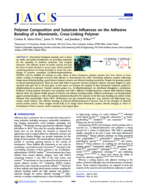
Hierarchical biological materials such as bone, sea shells, and marine bioadhesives are providing inspiration for the assembly of synthetic molecules into complex structures. The adhesive system of marine mussels has been the focus of much attention in recent years. Several catechol-containing polymers are being developed to mimic the cross-linking of proteins containing 3,4-dihydroxyphenylalanine (DOPA) used by shellfish for sticking to rocks. Many of these biomimetic polymer systems have been shown to form surface coatings or hydrogels; however, bulk adhesion is demonstrated less often. Developing adhesives requires addressing design issues including finding a good balance between cohesive and adhesive bonding interactions. Despite the growing number of mussel-mimicking polymers, there has been little effort to generate structure–property relations and gain insights on what chemical traits give rise to the best glues. In this report, we examine the simplest of these biomimetic polymers, poly[(3,4-dihydroxystyrene)-co-styrene]. Pendant catechol groups (i.e., 3,4-dihydroxystyrene) are distributed throughout a polystyrene backbone. Several polymer derivatives were prepared, each with a different 3,4-dihyroxystyrene content. Bulk adhesion testing showed where the optimal middle ground of cohesive and adhesive bonding resides. Adhesive performance was benchmarked against commercial glues as well as the genuine material produced by live mussels. In the best case, bonding was similar to that obtained with cyanoacrylate “Krazy Glue”. Performance was also examined using low- (e.g., plastics) and high-energy (e.g., metals, wood) surfaces. The adhesive bonding of poly[(3,4-dihydroxystyrene)-co-styrene] may be the strongest of reported mussel protein mimics. These insights should help us to design future biomimetic systems, thereby bringing us closer to development of bone cements, dental composites, and surgical glues.
DOI:
10.1021/ja303369p
Type of document:
Language:
Article
pubs.acs.org/JACS
Polymer Composition and Substrate Influences on the Adhesive
Bonding of a Biomimetic, Cross-Linking Polymer
Cristina R. Matos-Pérez,† James D. White,† and Jonathan J. Wilker*,†,‡
†
Department of Chemistry, Purdue University, 560 Oval Drive, West Lafayette, Indiana 47907-2084, United States
School of Materials Engineering, Purdue University, Neil Armstrong Hall of Engineering, 701 West Stadium Avenue, West Lafayette,
Indiana 47907-2045, United States
‡
ABSTRACT: Hierarchical biological materials such as bone,
sea shells, and marine bioadhesives are providing inspiration
for the assembly of synthetic molecules into complex
structures. The adhesive system of marine mussels has been
the focus of much attention in recent years. Several catecholcontaining polymers are being developed to mimic the crosslinking of proteins containing 3,4-dihydroxyphenylalanine
(DOPA) used by shellfish for sticking to rocks. Many of these biomimetic polymer systems have been shown to form
surface coatings or hydrogels; however, bulk adhesion is demonstrated less often. Developing adhesives requires addressing
design issues including finding a good balance between cohesive and adhesive bonding interactions. Despite the growing number
of mussel-mimicking polymers, there has been little effort to generate structure−property relations and gain insights on what
chemical traits give rise to the best glues. In this report, we examine the simplest of these biomimetic polymers, poly[(3,4dihydroxystyrene)-co-styrene]. Pendant catechol groups (i.e., 3,4-dihydroxystyrene) are distributed throughout a polystyrene
backbone. Several polymer derivatives were prepared, each with a different 3,4-dihyroxystyrene content. Bulk adhesion testing
showed where the optimal middle ground of cohesive and adhesive bonding resides. Adhesive performance was benchmarked
against commercial glues as well as the genuine material produced by live mussels. In the best case, bonding was similar to that
obtained with cyanoacrylate “Krazy Glue”. Performance was also examined using low- (e.g., plastics) and high-energy (e.g.,
metals, wood) surfaces. The adhesive bonding of poly[(3,4-dihydroxystyrene)-co-styrene] may be the strongest of reported
mussel protein mimics. These insights should help us to design future biomimetic systems, thereby bringing us closer to
development of bone cements, dental composites, and surgical glues.
■
to high-energy surfaces via metal chelation,14−18 individual
metal−ligand bonds,16,19 nonspecific adsorption,18 or hydrogen-bonding.18,20 Oxidative21,22 and enzymatic21−23 crosslinking may also be involved.
Incorporating DOPA and analogous reactive groups such as
catechol (i.e., 1,2-dihydroxybenzene) into polymers is being
pursued for a variety of applications. This field is expanding
rapidly, especially in the past 5 years, with many laboratories
contributing.24 Mussel mimetic polymers are being generated
from polypeptides,25−27 polyamides,28 polyacrylates,17,29−35
polyethylene glycols,36−52 polystyrenes,53−59 and polyurethanes.60 These polymers are enabling the development of
imaging agents,48 nanoparticle shells,44,48,61 elastomers,30,33,59
resins,58,62 coacervates,31 hydrogels,36−38,42,43 surface treatments,27,40,49,52 antibacterial coverings,51,63 and antifouling
coatings.34,35,45−47,50,51 A subset have shown the ability to
bond two substrates together.25,26,29−33,36−42,53,54,60
Whereas a coating requires only adhesive bonding to the
surface of interest, bulk glues also need the presence of cohesive
forces. These cohesive interactions are required to form the
majority of the material and reach between substrates to yield a
INTRODUCTION
Adhesives play a prominent role in everyday life, being used in
many industries including aerospace, automobile manufacturing, housing construction, wood products, packaging, and
labeling.1,2 Worldwide revenue generated by adhesives topped
$40 billion in 2010.3 New roles for specialty adhesives will be
found once we can develop the materials in demand for
applications such as surgical adhesives, orthopedic cements, and
dental glues. Marine biology can provide inspiration for the
design of such materials. The natural adhesive system of marine
mussels is receiving growing interest in the context of
biomimetics. These shellfish affix themselves to wet rocks by
assembling a cross-linked matrix of proteins.4,5 Essential to the
cross-linking chemistry of these proteins is the 3,4-dihydroxyphenylalanine (DOPA) residue.4,5 Several proteins have been
isolated from mussel adhesive plaques, each with DOPA
comprising between 3 and 30% of the total amino acid
content.4,5 A mechanism we have proposed for the formation of
mussel adhesive involves Fe3+ templating DOPA residues
followed by redox chemistry to generate radicals.6−13 Reactivity
of these radicals may bring about protein−protein coupling for
cohesive bonding within the bulk material and protein−
substrate linkages for surface adhesive bonding.12,13 Alternatively, or perhaps complementary, is direct binding of DOPA
© 2012 American Chemical Society
Received: April 8, 2012
Published: May 14, 2012
9498
dx.doi.org/10.1021/ja303369p | J. Am. Chem. Soc. 2012, 134, 9498−9505
Journal of the American Chemical Society
Article
dihydroxystyrene)-co-styrene] found lap shear bulk adhesion at
up to 1.2 ± 0.5 MPa.53 Over 1 MPa (∼145 pounds per square
inch (psi)) can be considered in the realm of high-strength
bonding and, once achieved, will enable development of
applications in several fields.1,2 Of course, even stronger
bonding is often desired.
Several factors influence the performance of an adhesive,
including the substrate type, surface preparation (e.g., roughness), cure conditions (e.g., temperature, time, humidity),
solvent, concentration, and viscosity.2 Beyond such formulation
issues, an appealing chemical aspect to explore is that of
polymer composition. By varying the ratio of 3,4-dihydroxystyrene:styrene within poly[(3,4-dihydroxystyrene)-co-styrene], we can gain access to a family of adhesive copolymers
with varied degrees of cross-linking. This type of systematic
study has not been carried out in detail with any other mussel
mimetic polymer system. Bonding performance described
below was examined on an array of low- to high-energy
surfaces: poly(tetrafluoroethylene) (PTFE, common name for
the DuPont product Teflon), poly(vinyl chloride) (PVC),
polished aluminum, sanded steel, and wood. Polymer
composition turns out to be a major factor dictating bonding
performance. This study presents the synthesis, characterization, and bulk adhesion of several polymers. We are excited
to report that the strongest bonding of these polymers displays
adhesion on par with that of commercial products such as
“Krazy Glue”, albeit with very different adhesion chemistry.
functional glue. Too much cohesion, however, will result in a
hardened material without significant affinity for a surface.
Likewise, too much adhesive bonding will come at the expense
of cohesion, and the bulk material will not exist. This balance of
cohesion and adhesion can be elusive, with no way to predict
where an optimal interplay may reside.
Despite the growing number of synthetic systems mimicking
aspects of mussel adhesive proteins, there have been few
detailed and systematic studies to illustrate which aspects of the
polymers give rise to the greatest bulk adhesion. In particular,
performance enhancements will arise from understanding how
the polymer composition dictates function. In other words:
How much pendant catechol should a polymer contain in order
to achieve the strongest bulk bonding? To answer this question,
we embarked upon a structure−property study in which the
relative contributions of cohesion and adhesion could be
changed systematically by altering the polymer composition.
The resulting insights will show where one might find the
highest-performing biomimetic material.
In an effort to gain straightforward chemical insights and also
to keep future scale-up in mind, our mimics of mussel adhesive
proteins are kept as simple as possible. The DOPA amino acid
can be stripped down to only a catechol group pendant from a
polymerizable olefin, hence the choice of 3,4-dihydroxystyrene
(Figure 1). To minimize structural and thermal perturbations
to the host polymer resulting from this monomer, polystyrene
was chosen to represent a protein backbone (Figure 1). Styrene
is commercially available and easy to polymerize on large scales.
A further advantage for these studies is that polystyrene alone
does not exhibit any appreciable bonding capability.53 The
target copolymer is thus poly[(3,4-dihydroxystyrene)-co-styrene], shown in Figure 1.
■
EXPERIMENTAL SECTION
Styrene and 3,4-dimethoxystyrene monomers were purchased and
purified with alumina columns for removal of polymerization
inhibitors. Details are provided in our earlier report.53 Solvents were
commercial anhydrous grade. A Varian Inova-300 MHz spectrometer
was used to collect NMR spectra. Gel permeation chromatography
(GPC) data were obtained using a Polymer Laboratories PL-GPC20
system and THF eluent. Polystyrene GPC standards (Varian, Inc.)
were used for instrument calibration. Differential scanning calorimetry
(DSC) data were obtained with a TA Instruments DSCQ2000
calorimeter.
Synthesis of Poly[(3,4-dimethoxystyrene)-co-styrene] Copolymers. In a typical polymerization, 2.86 mL (24.9 mmol) of
styrene and 3.70 mL (25.0 mmol) of 3,4-dimethoxystyrene were added
to a round-bottom flask with 30 mL of anhydrous toluene. The
reaction was cooled to −78 °C, and, after 10 min, 0.17 mL of nbutyllithium was added dropwise. The solution turned orange, was
stirred under an argon atmosphere for 8 h at −78 °C, and then was
allowed to warm to room temperature over 12 h of reaction.
Polymerization was quenched by addition of ∼1 mL of methanol.
Further addition of ∼100 mL of cold (−20 °C) methanol precipitated
the polymer. After isolation by filtering and drying under vacuum, at
least three rounds of dissolution in chloroform (∼15 mL) and
precipitation with methanol (∼100 mL) were used to remove
unreacted monomers. Yield of poly[(3,4-dimethoxystyrene)33-costyrene67] was 4.4 g, 33 mmol, 66%. 1H NMR (CDCl3): δ 0.6−2.3
ppm (broad, polymer backbone), 3.4−3.8 ppm (broad, methoxy
peaks), 6.0−7.4 ppm (broad, aromatic).
Synthesis of Poly[(3,4-dihydroxystyrene)-co-styrene]. Treatment with BBr3 and an acidic workup yielded the catechol-containing
polymers according to our previous methods.53 A typical deprotection
was accomplished by dissolving poly[(3,4-dimethoxystyrene)33%-costyrene67%] (4.4 g, 33 mmol) in 50.0 mL of anhydrous dichloromethane (DCM) under an argon atmosphere. The reaction was
cooled to 0 °C, and, after 10 min, BBr3 (1.2 mL, 13 mmol) was added
dropwise over 10 min. The solution was warmed to room temperature
and stirred overnight (∼12 h). The polymer was treated with 1% HCl
followed by an aqueous workup to obtain poly[(3,4-dihydroxystyrene)33%-co-styrene67%] (3.6 g, 27 mmol, 82%). Loss of the 1H NMR
Figure 1. Mussel adhesive is comprised of DOPA-containing proteins.
These proteins are mimicked with synthetic polymers by placing
pendant catechol groups along a polymer chain. One of the simplest
possible mimics is poly[(3,4-dihydroxystyrene)-co-styrene], in which
polystyrene represents the protein backbone and DOPA is represented
by 3,4-dihydroxystyrene.
Copolymers were prepared by a two-step synthetic route
developed in our laboratory previously.53 We have also made
cationic versions of these cross-linking polymers.54 Polymerization of styrene and 3,4-dimethoxystyrene yielded polymers
for which the ratio of monomers in the final polymers was
generally a reflection of the starting feed.53 The styrene and 3,4dihydroxystrene monomers distribute throughout the copolymer statistically or randomly, thereby providing a suitable
model for how DOPA residues are located within mussel
adhesive proteins.53 The relatively simple synthesis allows
access to large quantities of polymer, up to ∼20 g per reaction
in an academic laboratory. Our initial effort with poly[(3,49499
dx.doi.org/10.1021/ja303369p | J. Am. Chem. Soc. 2012, 134, 9498−9505
Journal of the American Chemical Society
Article
Table 1. Characterization Data for Poly[(3,4-dimethoxystyrene)-co-styrene] Copolymers
feed (%)
polymer observed (%)
3,4-dimethoxystyrene
styrene
3,4-dimethoxystyrene
styrene
Mn
Mw
PDI
Tg (°C)
0
5
9
15
22
50
50
51
53
100
95
91
85
78
50
50
49
47
0
5
10
15
19
26
33
42
36
100
95
90
85
81
74
67
58
64
32 300
37 500
39 800
40 700
40 900
49 600
57 500
50 575
32 700
38 400
48 800
50 000
48 700
54 500
65 800
84 200
61 700
43 800
1.2
1.3
1.2
1.2
1.3
1.3
1.5
1.2
1.3
106
103
100
93
97
67
62
60
68
methoxy peaks indicated complete deprotection. 1H NMR (CDCl3): δ
0.6−2.3 ppm (broad, polymer backbone) and 6.0−7.4 ppm (broad,
aromatic).
Adhesion Studies. Substrates for lap shear testing were prepared
by cutting each material into rectangular pieces, 8.89 cm long × 1.25
cm wide. A centered hole of 0.64 cm diameter was drilled into each
adherend 2.22 cm from one end. Aluminum was 0.318 cm thick, type
6061 T6, and mirror polished with Mibro no. 3 and Mibro no. 5 polish
followed by washing with hexanes, ethanol, acetone, and then
deionized water, 30 min each, and air-dried overnight. The steel
adherends, 0.318 cm thick, were sanded with 50 grit sandpaper prior
to testing and then washed with ethanol, acetone, and hexanes. PVC
(0.318 cm thick) and PTFE (0.953 cm thick) were obtained from
Ridout Plastics (San Diego, CA).
Red oak was purchased at a local hardware store and, after cutting
to 1.27 cm thick, had a surface roughness approximately equivalent to
that of 220 grit sandpaper. The wood adherends were cut and
adhesion strength was measured parallel to the wood grain, running
along the 8.89 cm edge of the adherend. Water loss from these wood
substrates may have occurred during the adhesive cure. Massing of
several oak adherends before versus after a typical cure treatment of 1
h at room temperature, 22 h at 55 °C, and 1 h at room temperature
revealed an average 4.12% decrease (e.g., from 10.1 to 9.68 g).
Lap shear adhesion measurements were conducted on an Instron
5544 materials testing system equipped with a 2000 N load cell.
Copolymer solutions in 1:1 acetone/DCM (0.3 g/mL, 22.5 μL) were
added to each adherend. Next, 15 μL of cross-linking solution (or
solvent when not adding the cross-linker) was added to deliver 0.33
equiv of cross-linker per catechol group. The adherends were
overlapped at 1.25 × 1.25 cm in a lap shear configuration (Figure
2). Each assembly was allowed to cure for 1 h at room temperature, 22
h at 55 °C, and then 1 h cooling at room temperature.
Figure 2 shows a representative extension versus force plot used for
quantifying adhesion. The early region of the trace is flat while the
crosshead moves up to begin loading the sample. Once the bond
begins to be stressed, a rise is seen until the sudden drop, indicating
bond breakage. Adherends were pulled apart at a rate of 2 mm/min.
The maximum bonding force in Newtons was recorded. Final adhesive
force in megapascals was obtained by dividing the maximum load at
failure, in Newtons, by the measured area of adhesive overlap in square
meters. For the polymer composition studies in Figure 3, each sample
was tested a minimum of 20 times, averaged, and reported with error
bars showing ±1 standard deviation. The comparisons to commercial
adhesives in Tables 2 and 3 were each tested a minimum of 10 times,
averaged, and reported with error bars showing ±1 standard deviation.
Tensile adhesion tests were carried out in an analogous manner using
aluminum rods of 1 cm diameter.
dimethoxystyrene content of each final polymer was similar
to that placed in the feed. Table 1 provides mole percent data
for each monomer in the feed versus that found in the isolated
polymers. For targeting low catechol polymers (e.g., 5.7a
0.5 ± 0.1
>5.7a
3.8 ± 0.7
0.7
0.4
0.36
1.5
0.7
0.2
0.1
0.04
0.3
0.1
polished aluminum
4
7
4
7
11
±
±
±
±
±
1
1
1
1
2
sanded steel
red oak
±
±
±
±
±
5.1 ± 0.9
10 ± 1
5±2
>10b
4±2
6
5
5.5
10
9
2
1
0.9
2
1
Substrate failed while adhesive bond remained intact. bExceeded range of the instrument.
conditions, concentration, added filler, viscosity, and addition of
adhesion promoters have all been examined. By contrast,
poly[(3,4-dihydroxystyrene)33%-co-styrene67%] is a relative newborn and, within the scope of this academic study, already
performs comparably to commercial products. Ideally, an
adhesive should be tailored for a target substrate. The
poly[(3,4-dihydroxystyrene)-co-styrene] with the strongest
bulk adhesion on aluminum is not necessarily the best polymer
for other substrates. Beyond changing polymers for each
surface, a detailed series of formulation efforts may enhance
performance even further.
Comparisons to Other Biomimetic Adhesive Polymers. We wished to place the performance of poly[(3,4dihydroxystyrene)33%-co-styrene67%] system within the expanding scope of other polymeric mussel protein mimics. Many of
these new systems are being used most often to generate
coatings27,34,35,40,45−47,49−52,63 or hydrogels,36−38,42,43 among
several other end goals, and some have shown adhesion.25,26,29−33,36−42,53,54,60 Direct comparisons of adhesive
performance are difficult to make given how many variables
are present including test methods, substrate composition,
surface preparations, solvents, viscosity, cure time, cure
temperature, and the presence or absence of water, among
several other conditions. Some of the stronger mussel mimics
reported are a polyurethane at 5.2 MPa,60 polypeptides
bonding up to 4.7 MPa,25 and a poly(ethylene glycol)/
polyacrylate at 1.2 MPa.29 Fusion proteins have been expressed
and modified to contain DOPA.76,77 These representations of
mussel proteins can adhere up to 4 MPa.67,78 The data in Table
3 indicate that poly[(3,4-dihydroxystyrene)33%-co-styrene67%] is
the strongest bonding synthetic mimic of mussel adhesive
tested to date. Maximum adhesion was at 10 ± 1 MPa for the
cross-linked polymer joining wood. Polished aluminum, sanded
steel, and PVC were adhered at greater than 5.7 MPa, also
stronger than that reported for other biomimetic adhesives.
Adhesion Strength of Synthetic Mimics Compared to
Plaques from Live Mussels. Recently we developed a
method for quantifying adhesive performance of the glue
produced by live mussels.79 On aluminum these shellfish
adhere at 0.3 ± 0.1 MPa.79 The byssal adhesive system of
mussels is comprised of plaques contacting the surface and
threads connecting each plaque to the animal’s soft inner body
(Figure 1). Tensile measurements were required to obtain
accurate adhesion data for the byssus. We were curious to see
how the performance of our biomimetic polymers compared to
the “real” material produced by mussels.
The polymer lap shear data from above (cf. Table 3) cannot
be compared directly to tensile measurements. In a lap shear
test, the substrates are overlapped and force is applied parallel
to the adhesive bond (Figure 2). Tensile testing is an end-toend butt joint, and the applied force is perpendicular to the
glue. Consequently, we gathered tensile adhesive data for
poly[(3,4-dihydroxystyrene)33%-co-styrene67%] on aluminum
rods. Pairs of tensile substrates were bonded together using
13.5 mg of dissolved poly[(3,4-dihydroxystyrene)33%-co-styrene67%] and (IO4)− over the 1 cm diameter overlap area.
Testing revealed that the polymeric adhesive was so strong that
not all of the joined substrates could be broken within the 2000
N capacity of our materials testing system. Some bonded
substrates pairs did separate and provided a lower limit of ≥9
MPa for poly[(3,4-dihydroxystyrene)33%-co-styrene67%] on
aluminum in tensile mode. The biomimetic system appears to
bond with significantly greater force than the natural material
after which the polymer was designed. Although we may be
trying to make the strongest possible glue, mussels need only
adhere as strongly as their environmental conditions dictate.
Indeed, if these shellfish were affixed to rocks any more
strongly, detachment forces exerted by waves or predators
might pull on the byssus to the point of damaging the soft,
internal tissues to which the threads connect (Figure 1).
■
CONCLUSIONS
With their ability to remain affixed to rocks in the turbulent
intertidal zone it is no wonder that mussels have inspired so
much research. Here we have presented a structure−property
study on the simplest mimic of mussel adhesive proteins.
Several copolymers were synthesized, characterized, and
examined for adhesive properties. A systematic approach was
taken in order to determine the polymer composition giving
rise to the greatest bulk adhesion. With the cross-linking and
surface bonding chemistries present in these copolymers, the
strongest adhesive is likely to provide a balance between
cohesive and adhesive bonding. Adhesion was quantified on
substrates ranging from low-energy, smooth plastics to highenergy, roughened metal. Performance was benchmarked
against common commercial glues as well as the native
material produced by live mussels. Adhesive performance of the
biomimetic polymer was comparable, and in some cases better,
than commercial products and the plaques of living shellfish.
Relative to other mussel mimetic polymers, poly[(3,4dihydroxystyrene)-co-styrene] appears to be the strongest
bulk adhesive. These comparisons, although interesting, are
difficult to make directly, given the broad variations in
conditions. Overall, these results help attest to the value of
using blueprints from biology when designing new materials.
Such a biomimetic approach may aid development of the
adhesives needed for industrial or biomedical applications
including wood glues without toxic formaldehyde, surgical
reattachment of soft tissues, and cements for connecting metal
implants to bone.
■
AUTHOR INFORMATION
Corresponding Author
wilker@purdue.edu
9503
dx.doi.org/10.1021/ja303369p | J. Am. Chem. Soc. 2012, 134, 9498−9505
Journal of the American Chemical Society
Article
Notes
(32) Shao, H.; Stewart, R. J. Adv. Mater. 2010, 22, 729−733.
(33) Chung, H. Y.; Glass, P.; Pothen, J. M.; Sitti, M.; Washburn, N.
R. Biomacromolecules 2011, 12, 342−347.
(34) Gao, C. L.; Li, G. Z.; Xue, H.; Yang, W.; Zhang, F. B.; Jiang, S.
Y. Biomaterials 2010, 31, 1486−1492.
(35) Li, G. Z.; Xue, H.; Cheng, G.; Chen, S. F.; Zhang, F. B.; Jiang, S.
Y. J. Phys. Chem. B 2008, 112, 15269−15274.
(36) Hu, B.; Messersmith, P. B. Orthod. Craniofacial Res. 2005, 8,
145−149.
(37) Lee, B. P.; Chao, C. Y.; Nunalee, F. N.; Motan, E.; Shull, K. R.;
Messersmith, P. B. Macromolecules 2006, 39, 1740−1748.
(38) Burke, S. A.; Ritter-Jones, M.; Lee, B. P.; Messersmith, P. B.
Biomed. Mater. 2007, 2, 203−210.
(39) Brubaker, C. E.; Kissler, H.; Wang, L.-J.; Kaufman, D. B.;
Messersmith, P. B. Biomaterials 2010, 31, 420−427.
(40) Murphy, J. L.; Vollenweider, L.; Xu, F. M.; Lee, B. P.
Biomacromolecules 2010, 11, 2976−2984.
(41) Brodie, M.; Vollenweider, L.; Murphy, J. L.; Xu, F. M.; Lyman,
A.; Lew, W. D.; Lee, B. P. Biomed. Mater. 2011, 6, 015014.
(42) Ryu, J. H.; Lee, Y.; Kong, W. H.; Kim, T. G.; Park, T. G.; Lee, H.
Biomacromolecules 2011, 12, 2653−2659.
(43) Holten-Andersen, N.; Harrington, M. J.; Birkedal, H.; Lee, B. P.;
Messersmith, P. B.; Lee, K. Y. C.; Waite, J. J. Proc. Natl. Acad. Sci.
U.S.A. 2011, 108, 2651−2655.
(44) Black, K. C. L.; Liu, Z. Q.; Messersmith, P. B. Chem. Mater.
2011, 23, 1130−1135.
(45) Finlay, A. S.; Dalsin, J.; Callow, M.; Callow, J. A.; Messersmith,
P. B. Biofouling 2006, 22, 391−399.
(46) Dalsin, J. L.; Lin, L. J.; Tosatti, S.; Voros, J.; Textor, M.;
Messersmith, P. B. Langmuir 2005, 21, 640−646.
(47) Lee, H.; Lee, K. D.; Pyo, K. B.; Park, S. Y.; Lee, H. Langmuir
2010, 26, 3790−3793.
(48) Bae, K. H.; Kim, Y. B.; Lee, Y.; Hwang, J.; Park, H.; Park, T. G.
Bioconjugate Chem. 2010, 21, 505−512.
(49) Chawla, K.; Lee, S.; Lee, B. P.; Dalsin, J. L.; Messersmith, P. B.;
Spencer, N. D. J. Biomed. Mater. Res. A 2009, 90A, 742−749.
(50) Pechey, A.; Elwood, C. N.; Wignall, G. R.; Dalsin, J. L.; Lee, B.
P.; Vanjecek, M.; Welch, I.; Ko, R.; Razvi, H.; Cadieux, P. A. J. Urology
2009, 182, 1628−1636.
(51) Yuan, S.; Wan, D.; Liang, B.; Pehkonen, S. O.; Ting, Y. P.;
Neoh, K. G.; Kang, E. T. Langmuir 2011, 27, 2761−2774.
(52) Malisova, B.; Tosatti, S.; Textor, M.; Gademann, K.; Zurcher, S.
Langmuir 2010, 26, 4018−4026.
(53) Westwood, G.; Horton, T. N.; Wilker, J. J. Macromolecules 2007,
40, 3960−3964.
(54) White, J. D.; Wilker, J. J. Macromolecules 2011, 44, 5085−5088.
(55) Yang, Z.; Pelton, R. Macromol. Rapid Commun. 1998, 19, 241−
246.
(56) Daly, W. H.; Moulay, S. J. Polym. Sci. 1986, 74, 227−242.
(57) Cristescu, R.; Mihailescu, I. N.; Stamatin, I.; Doraiswamy, A.;
Narayan, R.; Westwood, G.; Wilker, J. J.; Stafslien, S.; Chisholm, B.;
Chrisey, D. B. Appl. Surf. Sci. 2009, 255, 5496−5498.
(58) Bernard, J.; Branger, C.; Beurroies, I.; Denoyel, R.; Margaillan,
A. React. Funct. Polym. 2012, 72, 98−106.
(59) Pan, X. D.; Qin, Z. Q.; Yan, Y. Y.; Sadhukhan, O. Polymer 2010,
51, 3453−3461.
(60) Peiyu, S.; Liying, T.; Zhen, Z.; Xinling, W. Acta Polym. Sin. 2009,
803−808.
(61) Adkins, C. T.; Dobish, J. N.; Brown, C. S.; Mayrsohn, B.;
Hamilton, S. K.; Udoji, F.; Radford, K.; Yankeelov, T. E.; Gore, J. C.;
Harth, E. Polym. Chem. 2012, 3, 390−398.
(62) Kaneko, D.; Wang, S. Q.; Matsumoto, K.; Kinugawa, S.; Yasaki,
K.; Chi, D. H.; Kaneko, T. Polym. J. 2011, 43, 855−858.
(63) Han, H.; Wu, J.; Avery, C. W.; Mizutani, M.; Jiang, X.;
Kamigaito, M.; Chen, Z.; Xi, C.; Kuroda, K. Langmuir 2011, 27, 4010−
4019.
(64) Li, C.-H.; Chang, T.-C. Eur. Polym. J. 1991, 27, 35−39.
(65) Zhang, H.; Hong, K.; Mays, J. W. Macromolecules 2002, 25,
5738−5741.
The authors declare no competing financial interest.
■
ACKNOWLEDGMENTS
This work was supported by the Office of Naval Research, the
National Science Foundation, and a Ruth L. Kirschstein
National Research Service Award from the National Institute
of Health to C.R.M-P. We thank Harold McCarron and Jeffrey
Youngblood for insightful discussions, Kaumba Sakavuyi and
Matt Walters for obtaining DSC data, Allison Mattes for
assistance with GPC analysis, and Courtney Jenkins and
Heather Meredith for help collecting the commercial adhesion
data.
■
REFERENCES
(1) Pocius, A. V. Adhesion and Adhesives Technology. An Introduction,
2nd ed.; Carl Hanser Verlag: Munich, 2002.
(2) Petrie, E. M. Handbook of Adhesives and Sealants; McGraw Hill:
New York, 2007.
(3) Croson, M. E. Adhesives Sealants Ind. 2011, 18, 17−18.
(4) Waite, J. H. Integr. Comp. Biol. 2002, 42, 1172−1180.
(5) Sagert, J.; Sun, C.; Waite, J. H. In Biological Adhesives; Smith, A.
M., Callow, J. A., Eds.; Springer-Verlag: Berlin, 2006; pp 125−143.
(6) Hight, L. M.; Wilker, J. J. J. Mater. Sci. 2007, 42, 8934−8942.
(7) Loizou, E.; Weisser, J. T.; Dundigalla, A.; Porcar, L.; Schmidt, G.;
Wilker, J. J. Macromol. Biosci. 2006, 6, 711−718.
(8) Monahan, J.; Wilker, J. J. Chem. Commun. 2003, 1672−1673.
(9) Monahan, J.; Wilker, J. J. Langmuir 2004, 20, 3724−3729.
(10) Sever, M. J.; Weisser, J. T.; Monahan, J.; Srinivasan, S.; Wilker, J.
J. Angew. Chem., Int. Ed. 2004, 43, 448−450.
(11) Weisser, J. T.; Nilges, M. J.; Sever, M. J.; Wilker, J. J. Inorg.
Chem. 2006, 45, 7736−7747.
(12) Wilker, J. J. Curr. Opin. Chem. Biol. 2010, 14, 276−283.
(13) Wilker, J. J. Angew. Chem., Int. Ed. 2010, 49, 8076−8078.
(14) Lee, H.; Scherer, N. F.; Messersmith, P. B. Proc. Natl. Acad. Sci.
U.S.A. 2006, 103, 12999−13003.
(15) Ooka, A. A.; Garrell, R. L. Biopolymers (Biospectroscopy) 2000,
57, 92−102.
(16) Jankovic, I. A.; Saponjic, Z. V.; Comor, M. I.; Nedeljkovic, J. M.
J. Phys. Chem. C 2009, 113, 12645−12652.
(17) Wang, J. J.; Tahir, M. N.; Kappl, M.; Tremel, W.; Metz, N.; Barz,
M.; Theato, P.; Butt, H. J. Adv. Mater. 2008, 20, 3872−3876.
(18) Lana-Villarreal, T.; Rodes, A.; Pérez, J. M.; Gómez, R. J. Am.
Chem. Soc. 2005, 127, 12601−12611.
(19) McBride, M. B.; Wesselink, L. G. Environ. Sci. Technol. 1988, 22,
703−708.
(20) Mian, S. A.; Saha, L. C.; Jang, J.; Wang, L.; Gao, X.; Nagase, S. J.
Phys. Chem. C 2010, 114, 20793−20800.
(21) Fant, C.; Sott, K.; Elwing, H.; Hook, F. Biofouling 2000, 16,
119−132.
(22) Suci, P. A.; Geesey, G. G. J. Colloid Interface Sci. 2000, 230,
340−348.
(23) Burzio, L. A.; Waite, J. H. Biochemistry 2000, 39, 11147−11153.
(24) Moulay, S. C. R. Chim. 2009, 12, 577−601.
(25) Yu, M.; Deming, T. J. Macromolecules 1998, 31, 4739−4745.
(26) Wang, J.; Liu, C.; Lu, X.; Yin, M. Biomaterials 2007, 28, 3456−
3468.
(27) Saxer, S.; Portmann, C.; Tosatti, S.; Gademann, K.; Zurcher, S.;
Textor, M. Macromolecules 2010, 43, 1050−1060.
(28) Li, L.; Li, Y.; Luo, X. F.; Deng, J. P.; Yang, W. T. React. Funct.
Polym. 2010, 70, 938−943.
(29) Sarbjit, K.; Weerasekare, G. M.; Stewart, R. J. ACS Appl. Mater.
Interfaces 2011, 3, 941−944.
(30) Glass, P.; Chung, H. Y.; Washburn, N. R. Langmuir 2009, 25,
6607−6612.
(31) Shao, H.; Bachus, K. N.; Stewart, R. J. Macromol. Biosci. 2009, 9,
464−471.
9504
dx.doi.org/10.1021/ja303369p | J. Am. Chem. Soc. 2012, 134, 9498−9505
Journal of the American Chemical Society
Article
(66) Doraiswamy, A.; Dunaway, T. M.; Wilker, J. J.; Narayan, R. J. J.
Biomed. Mater. Res. B: Appl. Mater. 2009, 89B, 28−35.
(67) Cha, H. J.; Hwang, D. S.; Lim, S.; White, J. D.; Matos-Pérez, C.
R.; Wilker, J. J. Biofouling 2009, 25, 99−107.
(68) Zeng, H.; Hwang, D. S.; Israelachvili, J. N.; Waite, J. H. Proc.
Natl. Acad. Sci. U.S.A. 2010, 107, 12850−12853.
(69) Harrington, M. J.; Masic, A.; Holten-Andersen, N.; Waite, J. H.;
Fratzl, P. Science 2010, 328, 216−220.
(70) Sun, C.; Waite, J. H. J. Biol. Chem. 2005, 280, 39332−39336.
(71) Rzepecki, L. M.; Waite, J. H. Mol. Mar. Biol. Biotech. 1995, 4,
313−322.
(72) Papov, V. V.; Diamond, T. V.; Biemann, K.; Waite, J. H. J. Biol.
Chem. 1995, 270, 20183−20192.
(73) Vreeland, V.; Waite, J. H.; Epstein, L. J. Phycol. 1998, 34, 1−8.
(74) Zhao, H.; Waite, H. H. J. Biol. Chem. 2006, 281, 26150−26158.
(75) Rzepecki, L. M.; Hansen, K. M.; Waite, J. H. Biol. Bull. 1992,
183, 123−137.
(76) Hwang, D. S.; Gim, Y.; Cha, H. J. Biotechnol. Prog. 2005, 21,
965−970.
(77) Hwang, D. S.; Gim, Y.; Yoo, H. J.; Cha, H. J. Biomaterials 2007,
28, 3560−3568.
(78) Lim, S.; Choi, Y. S.; Kang, D. G.; Song, Y. H.; Cha, H. J.
Biomaterials 2010, 31, 3715−3722.
(79) Burkett, J. R.; Wojtas, J. L.; Cloud, J. L.; Wilker, J. J. J. Adhes.
2009, 85, 601−615.
9505
dx.doi.org/10.1021/ja303369p | J. Am. Chem. Soc. 2012, 134, 9498−9505
Coments go here:
- Log in to post comments