Rapidly curable chitosan–PEG hydrogels as tissue adhesives for hemostasis and wound healing
Rapidly curable chitosan–PEG hydrogels as tissue adhesives for hemostasis and wound healing
Folder:
Journal:
Year:
Article keywords:
Abstract:
Chitosan–poly(ethylene glycol)–tyramine (CPT) hydrogels were rapidly formed in situ using horseradish peroxidase and hydrogen peroxide to explore their performance as efficient tissueadhesives. A poly(ethylene glycol) modified with tyramine was grafted onto a chitosan backbone to enhance the solubility of the chitosan and to crosslink into three-dimensional networks. The elastic modulus of the hydrogels could be controlled by changing the crosslinking conditions, and the mechanical strength influenced the tissue adhesiveness of the hydrogels. The hydrogels showed the adhesiveness ranging from 3- to 20-fold that of fibrin glue (Greenplast®). The hemostatic ability of the hydrogels was evaluated on the basis that bleeding from liver defects was significantly arrested by the combined effect of the adhesiveness of the hydrogels and the hemostatic property of the chitosan materials. The enzymatic crosslinking method enabled the water-soluble chitosan to rapidly form hydrogels within 5 s of an incision into the skin of rats. Histological results demonstrated that the CPT hydrogels showed superior healing effects in the skin incision when compared to suture, fibrin glue and cyanoacrylate. By 2 weeks post-implantation, the wound was completely recovered, with a newly formed dermis, due to the presence of the CPT hydrogels in the incision. These results suggest that the in situ curablechitosanhydrogels are very interesting and promising tissueadhesive devices for biomedical applications.
DOI:
10.1016/j.actbio.2012.05.001
Type of document:
Language:
Acta Biomaterialia 8 (2012) 3261–3269
Contents lists available at SciVerse ScienceDirect
Acta Biomaterialia
journal homepage: www.elsevier.com/locate/actabiomat
Rapidly curable chitosan–PEG hydrogels as tissue adhesives for hemostasis
and wound healing
Eugene Lih, Jung Seok Lee, Kyung Min Park, Ki Dong Park ⇑
Department of Molecular Science & Technology, Ajou University, 5 Woncheon, Yeongtong, Suwon 443-749, Republic of Korea
a r t i c l e
i n f o
Article history:
Received 10 January 2012
Received in revised form 23 April 2012
Accepted 1 May 2012
Available online 19 May 2012
Keywords:
Chitosan
Poly(ethylene glycol)
Hydrogel
Tissue adhesive
Hemostasis
a b s t r a c t
Chitosan–poly(ethylene glycol)–tyramine (CPT) hydrogels were rapidly formed in situ using horseradish
peroxidase and hydrogen peroxide to explore their performance as efficient tissue adhesives. A poly(ethylene glycol) modified with tyramine was grafted onto a chitosan backbone to enhance the solubility of
the chitosan and to crosslink into three-dimensional networks. The elastic modulus of the hydrogels
could be controlled by changing the crosslinking conditions, and the mechanical strength influenced
the tissue adhesiveness of the hydrogels. The hydrogels showed the adhesiveness ranging from 3- to
20-fold that of fibrin glue (GreenplastÒ). The hemostatic ability of the hydrogels was evaluated on the
basis that bleeding from liver defects was significantly arrested by the combined effect of the adhesiveness of the hydrogels and the hemostatic property of the chitosan materials. The enzymatic crosslinking
method enabled the water-soluble chitosan to rapidly form hydrogels within 5 s of an incision into the
skin of rats. Histological results demonstrated that the CPT hydrogels showed superior healing effects
in the skin incision when compared to suture, fibrin glue and cyanoacrylate. By 2 weeks post-implantation, the wound was completely recovered, with a newly formed dermis, due to the presence of the CPT
hydrogels in the incision. These results suggest that the in situ curable chitosan hydrogels are very interesting and promising tissue adhesive devices for biomedical applications.
Ó 2012 Acta Materialia Inc. Published by Elsevier Ltd. All rights reserved.
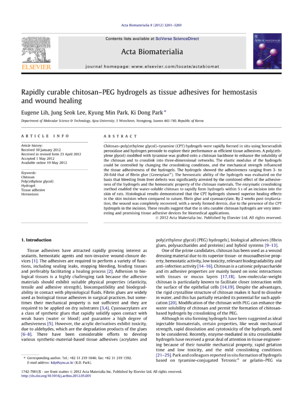
1. Introduction
Tissue adhesives have attracted rapidly growing interest as
sealants, hemostatic agents and non-invasive wound-closure devices [1]. The adhesives are required to perform a variety of functions, including sealing leaks, stopping bleeding, binding tissues
and preferably facilitating a healing process [2]. Adhesion to biological tissues is a highly challenging task because the adhesive
materials should exhibit suitable physical properties (elasticity,
tensile and adhesive strength), biocompatibility and biodegradability in contact with physiological fluids. Fibrin glues are widely
used as biological tissue adhesives in surgical practices, but sometimes their mechanical property is not sufficient and they are
required to be applied on dry substrates [3,4]. Cyanoacrylates are
a class of synthetic glues that rapidly solidify upon contact with
weak bases (water or blood) and guarantee a high degree of
adhesiveness [5]. However, the acrylic derivatives exhibit toxicity,
due to aldehydes, which are the degradation products of the glues
[6–8]. There have been considerable efforts to develop
various synthetic-material-based tissue adhesives (acrylates and
⇑ Corresponding author. Tel.: +82 31 219 1846; fax: +82 31 219 1592.
E-mail address: kdp@ajou.ac.kr (K.D. Park).
poly(ethylene glycol) (PEG) hydrogels), biological adhesives (fibrin
glues, polysaccharides and proteins) and hybrid systems [9–13].
One of the prime candidates, chitosan has been used as a wound
dressing material due to its superior tissue- or mucoadhesive property, hemostatic activity, low toxicity, relevant biodegradability and
anti-infection activity [14–16]. Chitosan is a cationic polysaccharide
and its adhesive properties are mainly based on ionic interactions
with tissues or mucus layers [17,18]. Low-molecular-weight
chitosan is particularly known to facilitate closer interaction with
the surface of the epithelial cells [14,19]. Despite the advantages,
the rigid crystalline structure of chitosan makes it hard to dissolve
in water, and this has partially retarded its potential for such application [20]. Modification of the chitosan with PEG can enhance the
water solubility of chitosan and permit the formation of chitosanbased hydrogels by crosslinking of the PEG.
Although in situ forming hydrogels have been suggested as ideal
injectable biomaterials, certain properties, like weak mechanical
strength, rapid dissolution and cytotoxicity of the hydrogels, need
to be considered. Recently, enzyme-mediated in situ crosslinkable
hydrogels have received a great deal of attention in tissue engineering because of their tunable mechanical property, rapid gelation
time and low toxicity, and the mild crosslinking conditions
[21–25]. Park and colleagues reported in situ formation of hydrogels
based on tyramine-conjugated TetronicÒ or gelatin–PEG via
1742-7061/$ - see front matter Ó 2012 Acta Materialia Inc. Published by Elsevier Ltd. All rights reserved.
http://dx.doi.org/10.1016/j.actbio.2012.05.001
3262
E. Lih et al. / Acta Biomaterialia 8 (2012) 3261–3269
Fig. 1. Schematic representation of in situ gel formation of CPT conjugates using HRP and H2O2.
enzymatic oxidative reactions using horseradish peroxidase (HRP)
and hydrogen peroxide (H2O2) [23–26]. HRP is a hemoprotein that
catalyzes the conjugation of phenol and aniline derivatives with
decomposed H2O2 molecules [27]. The enzymatically crosslinked
hydrogels showed excellent bioactivities and tunable physicochemical properties, suggesting that this type of hydrogel has great potential for use as an injectable material for tissue regenerative
medicine and various biomedical applications.
In this study, we report on enzyme-triggered in situ formation
of hydrogels based on chitosan as a tissue adhesive material for
hemostasis and wound healing. For the formation of the hydrogels,
chitosan was grafted with tyramine-modified PEGs and the tyramines were crosslinked by HRP and H2O2 as shown in Fig. 1. The
enzymatic crosslinking enabled the water-soluble chitosan to rapidly form hydrogels, which stably adhered to the wound site for a
desired period of time. The hydrogels were characterized in terms
of their physicomechanical properties, such as gelation time, elastic moduli and adhesive strengths, under various conditions. The
hemostatic and adhesive properties of the hydrogels as well as
the wound healing capability were also evaluated in vivo.
2. Materials and methods
2.1. Materials
Chitosan (low molecular weight, 75–85% deacetylated),
poly(ethylene glycol) (4000 g mol–1), HRP (units per mg solid
(using pyrogallol)), hydrogen peroxide, 4-dimethylamino pyridine
(DMAP) and p-nitrophenylchloroformate (PNC) were purchased
from Sigma–Aldrich (St. Louis, MO). Tyramine (TA) was purchased
from Acros Organics. Triethylamine (TEA) and aluminum oxide
were obtained from Kanto Chemical Co. and Strem Chemicals,
respectively. Fibrin glue kit (GreenplastÒ) was purchased from
Green Cross Co. and n-butyl-2-cyanoacrylate adhesive (HistoacrylÒ) was obtained from Tissueseal, LLC (Ann Arbor, MI, USA).
For cell culture, Dulbecco’s modified Eagle’s medium (high glucose), fetal bovine serum, trypsin/ethylenediaminetetraacetic acid,
penicillin–streptomycin and phosphate-buffered saline (PBS, pH
7.4) were obtained from Gibco BRL (Carlsbad, CA). Fluorescein
diacetate and ethidium bromide were purchased from Sigma–
aldrich. A Cell Counting Kit-8 was purchased from Dojindo (Kumamoto, Japan). All other chemicals and solvents were used such
without further purification.
2.2. Synthesis of chitosan–poly(ethylene glycol)–tyramine (CPT)
conjugates
The CPT conjugates were synthesized according to the previously
reported method [23]. Amine-reactive PEG (PNC–PEG–PNC) was
prepared by activating the hydroxyl groups of PEG with excess of
PNC. The activated PEG was reacted with TA and subsequently with
chitosan, to form the CPT conjugates (Fig. 2). Briefly, PEG (10 g,
5 mmol of hydroxyl groups) was dissolved in methylene chloride
(MC, 100 ml) at room temperature under a nitrogen atmosphere.
Fig. 2. Synthetic route of the CPT conjugates. Preparation of (a) PNC–PEG–PNC and (b) the conjugation of the PNC–PEG–TA with chitosan.
E. Lih et al. / Acta Biomaterialia 8 (2012) 3261–3269
A solution of DMAP (0.916 g, 7.5 mmol) and TEA (0.759 g, 7.5 mmol)
dissolved in MC (20 ml) was added to the PEG solution. The mixed
solution was stirred at room temperature for 15 min for the activation of the terminal hydroxyl groups. The mixture was then slowly
added to a solution of MC (50 ml) containing PNC (1.511 g,
7.5 mmol) in a dropwise manner. The reaction was carried out for
24 h at room temperature under N2. The resulting solution was
evaporated and precipitated in cold diethyl ether. The precipitate
was filtered and dried under vacuum to obtain the PNC–PEG–PNC
conjugates.
The CPT conjugates were prepared by coupling a chemically
modified PEG to a chitosan backbone. PNC–PEG–PNC (4 g, 2 mmol
of PNC groups) was dissolved in dimethyl sulfoxide (DMSO, 50 ml)
at room temperature under a nitrogen atmosphere. A solution of
TA (0.137 g, 1 mmol) dissolved in DMSO (25 ml) was added dropwise to the PNC–PEG–PNC solution, and reacted for 6 h under a
nitrogen atmosphere to prepare mono-TA-conjugated PEG (PNC–
PEG–TA). Chitosan (0.2 g) was dissolved in a co-solvent of dilute
hydrochloric acid solution (3 ml, pH 5) and DMSO (300 ml), and
the PNC–PEG–TA solution was added to the chitosan solution.
The mixture was stirred at room temperature under a nitrogen
atmosphere for 24 h. After completion of the reaction, the solution
was subjected to filtration using an aluminum oxide pad to remove
PNC salt, followed by dialysis (with a molecular weight cut-off of
12–14 kDa, SpectraPorÒ) against 0.01 M PBS solution (pH 7.4) for
3 days and then in distilled water for 2 days. The dialyzed solution
was lyophilized to obtain the CPT conjugates in the form of a white
powder.
The chemical structures of PNC–PEG–PNC and CPT were characterized by 1H nuclear magnetic resonance (NMR) spectroscopy
(Varian, 400 MHz spectrometer). The degree of substitution of
the TA groups was measured at a wavelength of 275 nm with
an ultraviolet (UV)–visible spectrometer (V-750 UV/VIS/NIR,
Jasco, Japan). The concentration of conjugated TA groups in the
CPT conjugates was calculated from the standard curve obtained
by monitoring the absorbance of a known concentration of TA
in deionized water. The chitosan and PEG compositions in the
CPT conjugates were evaluated by thermogravimetric analysis
(TGA), using a TGA Q50 analyzer (TA Instrument, USA). The
experiment was carried out under a nitrogen atmosphere with a
heating rate of 10 °C min–1 in the temperature range from 30 to
800 °C.
2.3. Gelation time measurements and rheological analysis
CPT hydrogels at a polymer concentration of 10 wt.% were dissolved in HRP solution (0.002–0.063 mg ml–1 of stock solution)
and H2O2 solution (0.06 wt.% of stock solution) in 0.01 M PBS (pH
7.4), and mixed under mild stirring. The gelation times of the
hydrogels were determined using the vial-tilting method [22].
The gel state was regarded as the condition when no flow was observed within a minute after inversion of the vial. The experiments
were performed in triplicate.
Rheological experiments (elastic modulus, G0 ) were carried out
with an Advanced Rheometer GEM-150–050 (Bohlin Instruments,
USA) using the parallel plates (20 mm diameter) configuration at
37 °C in oscillatory mode. The CPT polymers were dissolved both
in HRP solution (0.06 mg ml–1 of stock solution) and in solutions
containing different concentrations of H2O2 (0.015–0.06 wt.% of
stock solution). The polymer solutions containing HRP and H2O2
were rapidly mixed on the bottom plate of the rheometer and
the upper plate was immediately lowered down to a measuring
gap size of 1 mm. A frequency of 0.1 Hz (single frequency) and a
strain of 0.1% (strain control) were applied for the analysis to maintain the linear viscoelastic response.
3263
Fig. 3. Schematic illustration of the measurement of tissue adhesive strength.
2.4. Tissue adhesive strength
The adhesive properties were assessed by the following procedures, and are shown in Fig. 3. According to the method modified
from ASTM F2255–05 [28,29], the tissue adhesive strength of the
CPT hydrogels was measured using a universal testing machine
(Instron Model 3343, Norwood, MA, USA) at different H2O2 concentrations. Sections of porcine skin, used as substrates for the
experiment, were cut and cleaned to remove any excess fat. The
CPT solutions (10 wt.%) were dissolved in an HRP solution
(0.06 mg ml–1 of stock solution) and H2O2 solutions (0.015–
0.06 wt.% of stock solution). The HRP solution was applied on one
surface of the porcine skin (bonding area: 10 Â 10 mm2) and the
specimen was immediately covered with another tissue specimen
treated with the H2O2 solutions. During the test, the overlapped
skins were kept at room temperature for 30 min in a humid environment. The bonded skins were loaded until complete separation
was achieved at a crosshead speed of 10 mm min–1 with a 100 N
load cell. Measurements were performed on 20 samples of CPT
hydrogels in order to reduce the experimental error rate. Fibrin
glue was also measured under the same conditions as a control
adhesive (n = 5).
2.5. In vivo hemostatic ability test
To evaluate the hemostatic potential of the CPT hydrogels, a
hemorrhaging liver mouse model was employed (C57BL/6 mouse,
22–25 g, 5 weeks, male) [30–32]. All animal studies were performed in compliance with guidelines set by national regulations
and were approved by the local animal experiments ethical committee. Briefly, a mouse was anesthetized using zoletil and fixed
on a surgical corkboard. The liver of the mouse was exposed by
abdominal incision, and serous fluid around the liver was carefully
removed to prevent inaccuracies in the estimation of the blood
weight obtained by the filter paper. A pre-weighted filter paper
on a paraffin film was placed beneath the liver. Bleeding from
the liver was induced using a 20 G needle with the corkboard tilted
at about 30° and 50 ll of hydrogel was immediately applied to the
bleeding site using the dual syringe kit filled with the CPT solutions
(‘‘A’’ solution: 5 wt.% of CPT in an HRP solution (0.06 mg ml–1); ‘‘B’’
solution: 5 wt.% of CPT in a H2O2 solution (0.06 wt.%)). After 3 min,
the weight of the filter paper with absorbed blood was measured
and compared with a control group (no treatment after pricking
the liver).
2.6. Animal experiment for wound closure
All animal studies were performed in compliance with guidelines set by national regulations and were approved by the local
animal experiments ethical committee. To evaluate the bioadhesive property and the biocompatibility of the CPT hydrogels, rats
(normal SD rat, 100–150 g, 4 weeks, male) were anesthetized using
zoletil and their backs were shaved. Skin incisions 1.5 cm long and
skin thickness deep were made on both sides of the rat’s back [33].
The skin incisions were quickly closed by suture, fibrin glue, cyanoacrylate and CPT hydrogels. For this study, the CPT hydrogels were
3264
E. Lih et al. / Acta Biomaterialia 8 (2012) 3261–3269
prepared similarly to the hemostatic experiments. The hydrogels
were sterilized by filtration using 200 nm syringe filters and prepared with the dual syringe kit. A 50 ll aliquot of hydrogels was
applied to the wound area. At 7 and 14 days post-implantation,
the closure skin was harvested and fixed in p-formaldehyde solution (3.7 wt.%) for histological analysis by hematoxylin and eosin
(H&E) stain.
3. Results and discussion
3.1. Synthesis of CPT conjugates
The CPT conjugates were prepared by grafting PNCÀPEGÀTA
onto chitosan backbones. PNCÀPEGÀPNC was synthesized and
coupled with TA prior to the grafting. The PNC conjugation ratio
in the PEG chains was approximately 98% (molar ratio of PEG
and PNC $1:2), as previously reported [23]. The degree of TA conjugation with the PEG was calculated by 1H NMR spectroscopy, and
it was found that 97% of PEG in the CPT conjugates was functionalized with TA. The TA content in the CPT conjugates was also
determined by UV measurements (275 nm) and the TA content
was calculated to be $180 lmol g–1 of CPT (data not shown). The
weight ratio of chitosan and PEG in the CPT conjugates was determined by TGA (Fig. 4b). It was confirmed that the chitosan to PEG
ratio was 3:7 (w/w). The conjugate gave the following 1H NMR
peaks in D2O: d 1.86 (COCH3, chitosan), d 3.5–3.8 (ethylene groups
of PEG) and d 6.68 and 7.00 (aromatic protons of TA).
3.2. Rapid gelation time of CPT hydrogels
The CPT hydrogels were prepared by the simple blending of prefabricated polymer and enzyme solutions. In general, the in situ
preparation of the hydrogels takes about 5 s at an HRP concentration of 0.063 mg ml–1. A fast in situ gelation is essential in order to
quickly cover the defect surface and subsequently crosslink into
the hydrogels, which can adhere tightly to the bleeding surface
(typical gelation time of fibrin glues: 5 s). In contrast, very rapid
gelation can also result in the formation of non-homogeneous
hydrogels, which will result in insufficient mechanical properties
and adhesiveness. Notably, homogeneous and transparent CPT
hydrogels were formed under mild conditions, and no phase separation was observed. The gelation time of the CPT hydrogels could
be adjusted by changing the amount of the enzymes and the polymer concentration used for the preparation. Fig. 5 shows the gelation time of the CPT hydrogels with increasing concentrations of
CPT as a function of the amount of HRP. The HRP solutions containing CPT copolymer (10 wt.%) were mixed with H2O2 solution
(0.03 wt.%). The final concentrations of HRP were from 0.002 to
0.063 mg ml–1. A faster gelation time was obtained when more
HRP was involved in the crosslinking reactions, as the rate of the
Fig. 4. Characterizations of CPT conjugates: (a) 1H NMR spectrum and (b) TGA curves.
E. Lih et al. / Acta Biomaterialia 8 (2012) 3261–3269
3265
3.4. High tissue adhesive strength
Fig. 5. Gelation time of CPT hydrogels with respect to different CPT concentrations,
depending on the amount of HRP used for the reactions (0.002–0.063 mg ml–1)
(n = 5, mean ± SD).
formation of phenoxy radical was accelerated due to the high concentration of HRP. The radicals reacted with hydroxyl groups or
ortho-carbons of phenol groups in the CPT. The effect of CPT concentration on gelation time was significant at the lowest HRP concentration, but became negligible at higher concentrations.
3.3. Tunable mechanical property
The elastic moduli of the chitosan-based hydrogels were studied by oscillatory rheology experiments of polymer solutions containing HRP and H2O2 in PBS at 37 °C. The concentrations of the
CPT conjugates, H2O2, and HRP used in the formation of the CPT
hydrogels are listed in Table 1, and the change in the elastic modulus value of the CPT hydrogels is presented in Fig. 6. After mixing
the reactant solutions, there was a quick increase in the elastic
modulus in time due to the rapid enzymatic crosslinking reactions.
The plateau value reached was the G0 value, indicating the end of
the crosslinking process. The elastic modulus of the CPT hydrogels
could be controlled by changing the crosslinking conditions, e.g.
the concentration of H2O2. At an HRP concentration of
0.06 mg ml–1, the elastic modulus was about 8 kPa when
0.06 wt.% of H2O2 was used, and this value decreased with decreasing H2O2 concentration. This is because HRP catalyzes the conjugation of phenol groups with decomposed H2O2 molecules, and it is
assumed that H2O2 at less than 0.06 wt.% is not enough to fully
crosslink the hydrogels.
Fig. 6. Elastic moduli (G0 ) of CPT hydrogels (10 wt.%) depending on the different
concentrations of H2O2.
The adhesive strength of the CPT hydrogels was assessed at different H2O2 concentrations using the modified ASTM F2255–05
method, which is a standard test method for evaluating he strength
properties of tissue adhesives in lap-shear by tension loading
[28,29]. The CPT hydrogels were prepared using the same
conditions as used for the rheological studies (Table 1). Porcine
skins were used as the substrate materials (bonding area:
10 Â 10 mm2) and fibrin glue was used as the control bioadhesive.
None of the skin portions became unattached before the end of the
tests. When the tissue skins were sliding in different directions,
separation of two skins occurred due to the cohesion failure of
the hydrogels. This phenomenon could explain the relationship between the adhesive strength and the elastic modulus of the CPT
hydrogels. The hydrogels with an elastic modulus of 8 kPa showed
an adhesive strength of 97 kPa, whereas 17 kPa of adhesiveness
was obtained for the CPT hydrogels with an elastic modulus of
0.5 kPa. A higher mechanical strength led to a higher adhesive
strength. It is conceivable that the bulk property of the CPT hydrogels is correlated to the cohesive strength required to sustain a given amount of strain. In this case, the bonding between tissue and
adhesives could be relatively stable but the sample could be prone
to cohesive failure. This explanation is consistent with the previous
observations that fibrin sealants with a high elastic modulus failed
through an adhesive mode, while the fibrins with low stiffness primarily failed through a cohesive mode [34].
Interestingly, the CPT hydrogels exhibited adhesiveness ranging
from 3- to 20-fold that for the fibrin glue used in the study
(GreenplastÒ). The adhesive strength of the fibrin glue was about
5 kPa, whereas it was 17–97 kPa for the CPT hydrogels (Fig. 7).
The adhesive properties of chitosan have been attributed mainly
to the interaction between its positively charged amino groups
with negatively charged sialic acid groups present on the mucus
membrane [35]. It has also been reported that chitosan interacts
with the phospholipids of cell membranes mainly through electrostatic interactions, including hydrogen bonding and hydrophobic
forces, depending on the phospholipid packing density [36]. These
factors appear to affect the extent of the response to chitosan,
including the degree of acetylation and the molecular weight of
chitosan, which could facilitate closer interaction of chitosan with
the surface of epithelial cells [14]. Several reports have also shown
that chitosan and other cationic polymers are able to interact with
tight junctions in different epithelial cells, yielding a reversible
opening and reorientation of the tight junction without any permanent cell damage [14,19]. The general consensus regarding this
variable appears to be that a degree of deacetylation of more than
80% has the greatest effect on cells [19]. However, it is still not
Fig. 7. Adhesive strength of CPT hydrogels on porcine tissues (n = 20, mean ± SD).
The adhesiveness of fibrin glue was compared with that of the hydrogels.
3266
E. Lih et al. / Acta Biomaterialia 8 (2012) 3261–3269
Fig. 8. The evaluation of hemostatic ability of the hydrogel: (a) control and (b) CPT-D hydrogels. (c) Total blood loss from the damaged livers after 3 min. The black circle
denotes the CPT hydrogels sealing the liver (n = 3, mean ± SD).
Fig. 9. Photographs of wound closures. Skin incisions on back of the rats were treated by (a) suture, (b) fibrin glue, (c) CPT-D hydrogels and (d) cyanoacrylate.
clear how the intrinsic nature of chitosan imparts strong adhesive
properties at the molecular level.
Table 1
Concentrations of CPT conjugates, H2O2, and HRP used in the enzymatic crosslinking
reactions for the formation of CPT hydrogels.
CPT (wt.%)
3.5. Excellent hemostatic property of CPT hydrogels
Hemostatic agents are widely applied on hemorrhaging sites of
tissues or organs during intra-abdominal surgery [2,30,31]. Many
materials have been studied for their ability to arrest bleeding,
but they are not always effective in hemostasis; for example, the
agent should be in the liquid form before application and must rapidly solidify in the presence of body fluid with similar pliability as
of the damaged organs or tissues. In this study, CPT hydrogels were
explored as hemostatic tissue adhesives based on their previously
reported excellent adhesive properties [37]. Fig. 8a and b show
photographs of untreated bleeding liver and the extent of bleeding
after the application of hydrogels onto the liver, respectively. The
total blood loss from the control liver was about 154 mg for 3 min
after the liver was pricked with a needle. In contrast, the bleeding
was significantly arrested by the dressing of hydrogels, the loss of
blood being reduced to 59 mg through the combined effect of the
adhesiveness of the hydrogels and the hemostatic property of
chitosan (Fig. 8c). This result demonstrates that the CPT hydrogels
exhibit both elastic and adhesive properties when crosslinked
in situ, thus serving as an effective anti-hemorrhaging agent.
3.6. CPT hydrogels as wound closure materials
Recently, there has been an increase in the use of non-invasive
wound closure devices to avoid the pain of suturing and to lessen
the inconvenience caused to the patient [12,13]. Attractive alternatives to sutures and staples are required to rapidly adhere to the
skin close to the wound edges and to keep the wound closed for
a sufficient time, preferably with biodegradability, tissue regenerative property and minimal toxicity. To explore the use of CPT-D
hydrogels as wound closure devices, skin incisions on the backs
of rats were treated with CPT hydrogels and compared with
CPT-A
CPT-B
CPT-C
CPT-D
H2O2 (wt.%)
HRP (mg ml–1)
10
10
10
10
0.015
0.030
0.045
0.060
0.06
0.06
0.06
0.06
suture-, fibrin glue- and cyanoacrylate-treated models, as well as
normal skins. Fig. 9 presents photographs of the skin incisions just
after these treatments. Complete closure of the skin defect was observed for the suture and cyanoacrylate models. The CPT hydrogels
showed a small gap between the two edges of tissue, though the
size of the gap was less than in the case of the fibrin glue.
At 7 and 14 days after implantation, the skin samples were harvested and observed by H&E staining. Fig. 10 shows the H&Estained histology of cross-sections of normal skin and skin defects
sealed by suture, fibrin glue, cyanoacrylate and CPT-D hydrogel at
7 and 14 days post-wounding. In Fig. 10b, a large gap is apparent at
the incision site in the suture model and blood clots can be seen at
the point of application of the suture thread. After 14 days, the epidermal layer has penetrated into the sutured incision and the large
gap still remains, together with a suture hole (Fig. 10g). Cyanoacrylate performed much better with respect to wound closure than
suturing, although the gap was still present where the cyanoacrylate was applied (Fig. 10d). Fig. 10i reveals that the incision had
still not been completely crossed by newly formed collagen after
14 days, and the healing process was incomplete, demonstrating
fibrosis around the incision.
The fibrin glue-covered incision exhibited enhanced wound
healing when compared to the sutured and cyanoacrylate-covered
incisions at 14 days post-wounding. However, fibrosis was still observed, with incomplete dermis recovery. Compared with the other
three kinds of adhesive glue, CPT-D hydrogels showed superior
E. Lih et al. / Acta Biomaterialia 8 (2012) 3261–3269
3267
Fig. 10. H&E histological examination of CPT-D hydrogel (a–e) 7 and (f–j) 14 days after implantation, respectively; (a, f) normal tissue, (b, g) suture, (c, h) fibrin glue, (d, i)
cyanoacylate and (e, j) CPT-D hydrogel. Stratum corneum (w), epidermis (⁄), dermis (Â), and hair follicle (N) are presented. Size bars are 100 lm.
healing effects on the incision (Fig. 10e and j). The incision was
completely recovered, with a newly formed dermis and no visible
fibrosis, after 2 weeks. This observation can be explained by the
fact that chitosan improves the healing of the wound [14–16]. It
is known that chitosan evokes a minimal foreign body reaction,
with little or no fibrous encapsulation, acceleration of normal
3268
E. Lih et al. / Acta Biomaterialia 8 (2012) 3261–3269
granulation tissue formation, angiogenesis or rapid dermal regeneration [17,18]. Kim et al. reported that chitosan influenced all
stages of wound repair in experimental animal models [17].
Normally, chitosan-based hydrogels slowly degrade after several
months in vitro [38]. However, the rate of the degradation in wound
fluid would be much faster due to the secretion of overexpressed
and up-regulated lysozymes from the wound site. It has been
reported that the concentration of lysozyme in the wound fluid is
more than 0.5 mg ml–1 and the activity is 376 ± 240 U ml–1,
while it is even higher in the infected wound (4830 ± 1848 U ml–1)
[39–41]. For all materials, no sign of inflammation or infection in
the incisions was observed in this study.
4. Conclusions
Bioadhesive hydrogels based on chitosan were fabricated for
use as potential tissue adhesive materials. Enzyme-mediated
in situ crosslinking of hydrogels was performed using chitosan
grafted with PEG via HRP/H2O2. The mechanical strength as well
as the adhesiveness of the hydrogels was adjustable by using different concentrations of H2O2. This tunable adhesiveness makes
the hydrogels suitable as efficient bioadhesives in various medical
applications, which require a different range of adhesive strengths.
When applied to a rat liver defect or a rat skin incision model,
hydrogels cured by the enzymatic crosslinking method showed
excellent hemostatic properties and wound healing effects within
5 s. We propose that in situ curable chitosan hydrogels are useful
as bioadhesives for numerous medical applications. In a future
investigation, mechanistic studies on the strong tissue adhesiveness of the chitosan hydrogels will be performed in the molecular
level. Moreover, the in vivo degradation of hydrogels in various tissue defects will also be evaluated.
Acknowledgements
This work was supported by the National Research Foundation
of Korea (NRF) Grant funded by the Korea government (MEST)
(2010-0027776), Nano-Biotechnology Project (Regenomics), Ministry of Science & Technology (2011-0007746 (B020214)), and
the Korea Science and Engineering Foundation, Ministry of Education, Science and Technology (2011-0001805).
Appendix A. Figures with essential colour discrimination
Certain figures in this article, particularly Figs. 1, 3, 4, 8, 9 and
10, are difficult to interpret in black and white. The full colour
images can be found in the on-line version, at http://dx.doi.org/
10.1016/j.actbio.2012.05.001.
Appendix B. Supplementary data
Supplementary data associated with this article can be found, in
the online version, at http://dx.doi.org/10.1016/j.actbio.2012.05.
001.
References
[1] Reece TB, Maxey TS, Kron IL. A prospectus on tissue adhesives. Am J Surg
2001;182:S40–4.
[2] Spotnitz WD, Burks S. Hemostats, sealants, and adhesives: components of the
surgical toolbox. Transfusion 2008;48:1502–16.
[3] MacGillivray TE. Fibrin sealants and glues. J Card Surg 2003;18:480–5.
[4] Sierra DH. Fibrin sealant adhesive systems: a review of their chemistry,
material properties and clinical applications. J Biomater Appl 1993;7:309–52.
[5] Mobley SR, Hilinski J, Toriumi DM. Surgical tissue adhesives. Facial Plast Surg
Clin North Am 2002;10:147–54.
[6] Leggat PA, Smith DR, Kedjarune U. Surgical applications of cyanoacrylate
adhesives: a review of toxicity. ANZ J Surg 2007;77:209–13.
[7] Singer AJ, Quinn JV, Hollander JE. The cyanoacrylate topical skin adhesives. Am
J Emerg Med 2008;26:490–6.
[8] Eaglstein WH, Sullivan T. Cyanoacrylates for skin closure. Dermatol Clin
2005;23:193–8.
[9] Chen Q, Liang S, Thouas GA. Synthesis and characterisation of poly(glycerol
sebacate)-co-lactic acid as surgical sealants. Soft Matter 2011;7:6484–92.
[10] Chen T, Janjua R, McDermott MK, Bernstein SL, Steidl SM, Payne GF. Gelatinbased biomimetic tissue adhesive. Potential for retinal reattachment. J Biomed
Mater Res Part B: Appl Biomater 2006;77B:416–22.
[11] Lee Y, Chung HJ, Yeo S, Ahn C-H, Lee H, Messersmith PB, et al. Thermosensitive, injectable, and tissue adhesive sol–gel transition hyaluronic acid/
pluronic composite hydrogels prepared from bio-inspired catechol–thiol
reaction. Soft Matter 2010;6:977–83.
[12] Oelker AM, Grinstaff MW. Ophthalmic adhesives: a materials chemistry
perspective. J Mater Chem 2008;18:2521–36.
[13] Ryou M, Thompson CC. Tissue adhesives: a review. Tech Gastrointest Endosc
2006;8:33–7.
[14] Ibrahim AA. Chitosan topical gel formulation in the management of burn
wounds. Int J Biol Macromol 2009;45:16–21.
[15] Jayakumar R, Prabaharan M, Sudheesh Kumar PT, Nair SV, Tamura H.
Biomaterials based on chitin and chitosan in wound dressing applications.
Biotechnol Adv 2011;29:322–37.
[16] Ueno H, Mori T, Fujinaga T. Topical formulations and wound healing
applications of chitosan. Adv Drug Deliv Rev 2001;52:105–15.
[17] Kim I-Y, Seo S-J, Moon H-S, Yoo M-K, Park I-Y, Kim B-C, et al. Chitosan and its
derivatives for tissue engineering applications. Biotechnol Adv 2008;26:1–21.
˘
˘
[18] Sezer A, Hatipoglu F, Cevher E, Ogurtan Z, Bas A, Akbuga J. Chitosan film
containing fucoidan as a wound dressing for dermal burn healing: preparation
and in vitro/in vivo evaluation. AAPS Pharm Sci Tech 2007;8:E94–E101.
[19] Smith J, Wood E, Dornish M. Effect of chitosan on epithelial cell tight junctions.
Pharm Res 2004;21:43–9.
[20] Park JH, Cho YW, Chung H, Kwon IC, Jeong SY. Synthesis and characterization
of sugar-bearing chitosan derivatives: aqueous solubility and biodegradability.
Biomacromolecules 2003;4:1087–91.
[21] Jin R, Hiemstra C, Zhong Z, Feijen J. Enzyme-mediated fast in situ formation of
hydrogels
from
dextran–tyramine
conjugates.
Biomaterials
2007;28:2791–800.
[22] Kurisawa M, Chung JE, Yang YY, Gao SJ, Uyama H. Injectable biodegradable
hydrogels composed of hyaluronic acid-yramine conjugates for drug delivery
and tissue engineering. Chem Commun 2005;43:12–4.
[23] Park KM, Ko KS, Joung YK, Shin H, Park KD. In situ cross-linkable gelatin–
poly(ethylene glycol)–tyramine hydrogel via enzyme-mediated reaction for
tissue regenerative medicine. J Mater Chem 2011;21:13180–7.
[24] Park KM, Shin YM, Joung YK, Shin H, Park KD. In situ forming hydrogels based
on tyramine conjugated 4-arm-PPO–PEO via enzymatic oxidative reaction.
Biomacromolecules 2010;11:706–12.
[25] Tran NQ, Joung YK, Lih E, Park KD. In situ forming and rutin-releasing chitosan
hydrogels
as
injectable
dressings for
dermal
wound healing.
Biomacromolecules 2011;12:2872–80.
[26] Park KM, Jun I, Joung YK, Shin H, Park KD. In situ hydrogelation and RGD
conjugation of tyramine-conjugated 4-arm PPO–PEO block copolymer for
injectable bio-mimetic scaffolds. Soft Matter 2011;7:986–92.
[27] Lee F, Chung JE, Kurisawa M. An injectable hyaluronic acid–tyramine hydrogel
system for protein delivery. J Control Release 2009;134:186–93.
[28] Kull S, Martinelli I, Briganti E, Losi P, Spiller D, Tonlorenzi S, et al. Glubran2
surgical glue: in vitro evaluation of adhesive and mechanical properties. J Surg
Res 2009;157:e15–21.
[29] Ninan L, Monahan J, Stroshine RL, Wilker JJ, Shi R. Adhesive strength of marine
mussel extracts on porcine skin. Biomaterials 2003;24:4091–9.
[30] Liu Y, Kopelman D, Wu L-Q, Hijji K, Attar I, Preiss-Bloom O, et al. Biomimetic
sealant based on gelatin and microbial transglutaminase: an initial in vivo
investigation. J Biomed Mater Res Part B: Appl Biomater 2009;91B:5–16.
[31] Murakami Y, Yokoyama M, Nishida H, Tomizawa Y, Kurosawa H. In vivo and
in vitro evaluation of gelation and hemostatic properties of a novel tissueadhesive hydrogel containing a cross-linkable polymeric micelle. J Biomed
Mater Res Part B: Appl Biomater 2009;91B:102–8.
[32] Ryu JH, Lee Y, Kong WH, Kim TG, Park TG, Lee H. Catechol-functionalized
chitosan/pluronic hydrogels for tissue adhesives and hemostatic materials.
Biomacromolecules 2011;12:2653–9.
[33] Mo X, Iwata H, Ikada Y. A tissue adhesives evaluated in vitro and in vivo
analysis. J Biomed Mater Res Part A 2010;94A:326–32.
[34] Serrero Al, Trombotto Sp, Bayon Y, Gravagna P, Montanari S, David L.
Polysaccharide-based adhesive for biomedical applications: correlation
between
rheological
behavior
and
adhesion.
Biomacromolecules
2011;12:1556–66.
[35] Fiebrig I, Harding S, Davis S. Sedimentation analysis of potential interactions
between mucins and a putative bioadhesive polymer ultracentrifugation. Prog
Colloid Polym Sci 1994;94:66–73.
[36] Pavinatto FJ, Pavinatto A, Caseli L, dos Santos DS, Nobre TM, Zaniquelli MED,
et al. Interaction of chitosan with cell membrane models at the air–water
interface. Biomacromolecules 2007;8:1633–40.
[37] Samyn P, Ruhe Jr, Biesalski M. Polymerizable biomimetic vesicles with
controlled local presentation of adhesive functional DOPA groups. Langmuir
2010;26:8573–81.
[38] Shi C, Zhu Y, Ran X, Wang M, Su Y, Cheng T. Therapeutic potential of chitosan
and its derivatives in regenerative medicine. J Surg Res 2006;133:185–92.
E. Lih et al. / Acta Biomaterialia 8 (2012) 3261–3269
[39] Choi JS, Yoo HS. Pluronic/chitosan hydrogels containing epidermal growth
factor with wound-adhesive and photo-crosslinkable properties. J Biomed
Mater Res Part A 2010;95A:564–73.
[40] Frohm M, Gunne H, Bergman A-C, Agerberth B, Bergman T, Boman A, et al.
Biochemical and antibacterial analysis of human wound and blister fluid. Eur J
Biochem 1996;237:86–92.
3269
[41] Hasmann A, Wehrschuetz-Sigl E, Kanzler G, Gewessler U, Hulla E, Schneider
KP, et al. Novel peptidoglycan-based diagnostic devices for detection of wound
infection. Diagn Microbiol Infect Dis 2011;71:12–23.
Coments go here:
- Log in to post comments